| Entry | Database: PDB / ID: 7v63
|
|---|
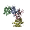
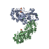
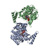
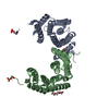
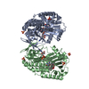
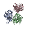
| Title | Structure of dimeric uPAR at low pH |
|---|
 Components Components | Urokinase plasminogen activator surface receptor |
|---|
 Keywords Keywords | IMMUNE SYSTEM / GPI-anchored protein |
|---|
| Function / homology |  Function and homology information Function and homology information
urokinase plasminogen activator receptor activity / Attachment of GPI anchor to uPAR / positive regulation of homotypic cell-cell adhesion / urokinase plasminogen activator signaling pathway / regulation of plasminogen activation / regulation of fibrinolysis / protein complex involved in cell-matrix adhesion / serine-type endopeptidase complex / Dissolution of Fibrin Clot / extrinsic component of membrane ...urokinase plasminogen activator receptor activity / Attachment of GPI anchor to uPAR / positive regulation of homotypic cell-cell adhesion / urokinase plasminogen activator signaling pathway / regulation of plasminogen activation / regulation of fibrinolysis / protein complex involved in cell-matrix adhesion / serine-type endopeptidase complex / Dissolution of Fibrin Clot / extrinsic component of membrane / positive regulation of DNA binding / positive regulation of epidermal growth factor receptor signaling pathway / negative regulation of intrinsic apoptotic signaling pathway / positive regulation of release of cytochrome c from mitochondria / regulation of proteolysis / regulation of cell adhesion / specific granule membrane / cell projection / positive regulation of protein phosphorylation / chemotaxis / blood coagulation / signaling receptor activity / endoplasmic reticulum lumen / signaling receptor binding / protein domain specific binding / external side of plasma membrane / focal adhesion / Neutrophil degranulation / endoplasmic reticulum membrane / negative regulation of apoptotic process / enzyme binding / cell surface / signal transduction / extracellular region / membrane / plasma membraneSimilarity search - Function CD59 antigen, conserved site / Ly-6 / u-PAR domain signature. / u-PAR/Ly-6 domain / Ly-6 antigen / uPA receptor -like domain / Ly-6 antigen/uPA receptor-like / Snake toxin-like superfamilySimilarity search - Domain/homology |
|---|
| Biological species |  Homo sapiens (human) Homo sapiens (human) |
|---|
| Method |  X-RAY DIFFRACTION / SYNCHROTRON / SAD / Resolution: 2.906 Å X-RAY DIFFRACTION / SYNCHROTRON / SAD / Resolution: 2.906 Å |
|---|
 Authors Authors | Yuan, C. / Huang, M. |
|---|
| Funding support |  China, 1items China, 1items | Organization | Grant number | Country |
|---|
| National Natural Science Foundation of China (NSFC) | 22077016, 82070142, 31570745, 31670739 | China |
|
|---|
 Citation Citation | Journal: Nat Commun / Year: 2022
Title: Crystal structure and cellular functions of uPAR dimer
Authors: Yu, S. / Sui, Y. / Wang, J. / Li, Y. / Li, H. / Cao, Y. / Chen, L. / Jiang, L. / Yuan, C. / Huang, M. |
|---|
| History | | Deposition | Aug 19, 2021 | Deposition site: PDBJ / Processing site: PDBJ |
|---|
| Revision 1.0 | Dec 22, 2021 | Provider: repository / Type: Initial release |
|---|
| Revision 1.1 | Apr 6, 2022 | Group: Database references / Category: citation / citation_author
Item: _citation.journal_volume / _citation.page_first ..._citation.journal_volume / _citation.page_first / _citation.pdbx_database_id_DOI / _citation.title / _citation.year |
|---|
| Revision 1.2 | Oct 30, 2024 | Group: Data collection / Structure summary
Category: chem_comp_atom / chem_comp_bond ...chem_comp_atom / chem_comp_bond / pdbx_entry_details / pdbx_modification_feature
Item: _pdbx_entry_details.has_protein_modification |
|---|
|
|---|
 Movie
Movie Controller


 Open data
 Basic information
 Structure visualization
 Downloads & links
 Other downloads


 PDBj
PDBj
 Assembly