 Movie
Movie Controller

[English] 日本語
 Yorodumi
- PDB-7v4m: Unique non-heme hydroxylase in biosynthesis of nucleoside antibio... -
+ Open data

- Basic information
| Entry | Database: PDB / ID: 7v4m | ||||||
|---|---|---|---|---|---|---|---|
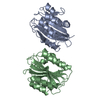
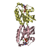
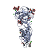
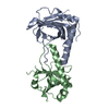
| Title | Unique non-heme hydroxylase in biosynthesis of nucleoside antibiotic pathway uncover mechanism of reaction | ||||||
 Components Components | Beta-hydroxylase | ||||||
 Keywords Keywords | ANTIBIOTIC / caprazamycin / MraY / beta-hydroxylase | ||||||
| Function / homology |  Function and homology information Function and homology information | ||||||
| Biological species |  Streptomyces sp. MK730-62F2 (bacteria) Streptomyces sp. MK730-62F2 (bacteria) | ||||||
| Method |  X-RAY DIFFRACTION / SYNCHROTRON / MOLECULAR REPLACEMENT / Resolution: 1.9 Å X-RAY DIFFRACTION / SYNCHROTRON / MOLECULAR REPLACEMENT / Resolution: 1.9 Å | ||||||
 Authors Authors | Li, T.L. / Saeid, M.Z. | ||||||
| Funding support |  Taiwan, 1items Taiwan, 1items
| ||||||
 Citation Citation | Journal: Commun Chem / Year: 2022 Title: beta-Hydroxylation of alpha-amino-beta-hydroxylbutanoyl-glycyluridine catalyzed by a nonheme hydroxylase ensures the maturation of caprazamycin Authors: Zadeh, S.M. / Chen, M.H. / Wang, Z.C. / Astani, E.K. / Lo, I.W. / Lin, K.H. / Hsu, N.S. / Adhikari, K. / Lyu, S.Y. / Tsai, H.Y. / Terasawa, Y. / Yabe, M. / Yamamoto, K. / Ichikawa, S. / Li, T.L. | ||||||
| History |
|
- Structure visualization
| Structure viewer | Molecule: MolmilJmol/JSmol |
|---|
- Downloads & links
-Download
| PDBx/mmCIF format | 7v4m.cif.gz | 55 KB | Display | PDBx/mmCIF format |
|---|---|---|---|---|
| PDB format | pdb7v4m.ent.gz | 36.1 KB | Display | PDB format |
| PDBx/mmJSON format | 7v4m.json.gz | Tree view | PDBx/mmJSON format | |
| Others |  Other downloads Other downloads |
-Validation report
| Arichive directory | https://data.pdbj.org/pub/pdb/validation_reports/v4/7v4mftp://data.pdbj.org/pub/pdb/validation_reports/v4/7v4m | HTTPS FTP |
|---|
-Related structure data
| Related structure data |  7v4fSC 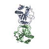 7v4nC 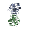 7v4oC 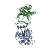 7v4pC S: Starting model for refinement C: citing same article ( |
|---|---|
| Similar structure data |


-Links
 PDBj
PDBj

- Assembly
| Deposited unit | 
| |||||||||
|---|---|---|---|---|---|---|---|---|---|---|
| 1 | 
| |||||||||
| Unit cell |
| |||||||||
| Components on special symmetry positions |
|
-Components
-Protein , 1 types, 1 molecules B
| #1: Protein | Mass: 21090.766 Da / Num. of mol.: 1 Source method: isolated from a genetically manipulated source Source: (gene. exp.) Streptomyces sp. MK730-62F2 (bacteria) / Gene: cpz10Production host: References: UniProt: C4NCJ7 |
|---|
-Non-polymers , 5 types, 140 molecules 


| #2: Chemical | ChemComp-AKG /  Mass: 146.098 Da / Num. of mol.: 1 / Source method: obtained synthetically / Formula: C5H6O5 Mass: 146.098 Da / Num. of mol.: 1 / Source method: obtained synthetically / Formula: C5H6O5 | ||||
|---|---|---|---|---|---|
| #3: Chemical | ChemComp-K /  Mass: 39.098 Da / Num. of mol.: 1 / Source method: obtained synthetically / Formula: K Mass: 39.098 Da / Num. of mol.: 1 / Source method: obtained synthetically / Formula: K | ||||
| #4: Chemical |  Mass: 55.845 Da / Num. of mol.: 2 / Source method: obtained synthetically / Formula: Fe Mass: 55.845 Da / Num. of mol.: 2 / Source method: obtained synthetically / Formula: Fe#5: Chemical | ChemComp-PO4 / |  Mass: 94.971 Da / Num. of mol.: 1 / Source method: obtained synthetically / Formula: PO4 Mass: 94.971 Da / Num. of mol.: 1 / Source method: obtained synthetically / Formula: PO4#6: Water | ChemComp-HOH / | Mass: 18.015 Da / Num. of mol.: 135 / Source method: isolated from a natural source / Formula: H2O |
-Details
| Has ligand of interest | Y |
|---|
-Experimental details
-Experiment
| Experiment | Method: X-RAY DIFFRACTION / Number of used crystals: 1 |
|---|
- Sample preparation
| Crystal | Density Matthews: 2.35 Å3/Da / Density % sol: 47.76 % |
|---|---|
| Crystal grow | Temperature: 293 K / Method: vapor diffusion, hanging drop Details: 0.1M Calcium acetate 0.1M MES pH 6.4 9% V/V PEG 4K 10 mg |
-Data collection
| Diffraction | Mean temperature: 80 K / Serial crystal experiment: N |
|---|---|
| Diffraction source | Source: SYNCHROTRON / Site: NSRRC / Beamline: BL15A1 / Wavelength: 0.9 Å |
| Detector | Type: RAYONIX MX300HE / Detector: CCD / Date: Apr 17, 2021 |
| Radiation | Protocol: SINGLE WAVELENGTH / Monochromatic (M) / Laue (L): M / Scattering type: x-ray |
| Radiation wavelength | Wavelength: 0.9 Å / Relative weight: 1 |
| Reflection | Resolution: 1.9→19.86 Å / Num. obs: 16089 / % possible obs: 100 % / Redundancy: 5.3 % / CC1/2: 0.78 / Net I/σ(I): 23.6 |
| Reflection shell | Resolution: 1.9→1.97 Å / Num. unique obs: 1596 / CC1/2: 0.78 |
- Processing
| Software |
| |||||||||||||||||||||||||||||||||||||||||||||||||||||||||||||||||||||||||||||
|---|---|---|---|---|---|---|---|---|---|---|---|---|---|---|---|---|---|---|---|---|---|---|---|---|---|---|---|---|---|---|---|---|---|---|---|---|---|---|---|---|---|---|---|---|---|---|---|---|---|---|---|---|---|---|---|---|---|---|---|---|---|---|---|---|---|---|---|---|---|---|---|---|---|---|---|---|---|---|
| Refinement | Method to determine structure: MOLECULAR REPLACEMENT Starting model: 7V4F Resolution: 1.9→19.86 Å / SU ML: 0.2 / Cross valid method: FREE R-VALUE / σ(F): 1.36 / Phase error: 22.13 / Stereochemistry target values: ML
| |||||||||||||||||||||||||||||||||||||||||||||||||||||||||||||||||||||||||||||
| Solvent computation | Shrinkage radii: 0.9 Å / VDW probe radii: 1.11 Å / Solvent model: FLAT BULK SOLVENT MODEL | |||||||||||||||||||||||||||||||||||||||||||||||||||||||||||||||||||||||||||||
| Refinement step | Cycle: LAST / Resolution: 1.9→19.86 Å
| |||||||||||||||||||||||||||||||||||||||||||||||||||||||||||||||||||||||||||||
| Refine LS restraints |
| |||||||||||||||||||||||||||||||||||||||||||||||||||||||||||||||||||||||||||||
| LS refinement shell |
|
