 Movie
Movie Controller


+ Open data

- Basic information
| Entry | Database: PDB / ID: 7v3q | ||||||
|---|---|---|---|---|---|---|---|
| Title | Crystal structure of anti-MUC1 antibody 16A | ||||||
 Components Components |
| ||||||
 Keywords Keywords | IMMUNE SYSTEM / Antibody / anti-MUC1 / Cancer / ANTITUMOR PROTEIN | ||||||
| Function / homology | Immunoglobulins / Immunoglobulin-like / Sandwich / Mainly Beta Function and homology information Function and homology information | ||||||
| Biological species |  | ||||||
| Method |  X-RAY DIFFRACTION / MOLECULAR REPLACEMENT / Resolution: 2.98 Å X-RAY DIFFRACTION / MOLECULAR REPLACEMENT / Resolution: 2.98 Å | ||||||
 Authors Authors | Niu, J. / Xu, L. / Meng, B. / Han, Y.B. / Yang, B. | ||||||
| Funding support |  China, 1items China, 1items
| ||||||
 Citation Citation | Journal: To Be Published Title: Site-specific GalNAc modification on a MUC1 neoantigen epitope forms a basis for high-affinity antibody binding Authors: Han, Y.B. / Xu, L. | ||||||
| History |
|
- Structure visualization
| Structure viewer | Molecule: MolmilJmol/JSmol |
|---|
- Downloads & links
-Download
| PDBx/mmCIF format | 7v3q.cif.gz | 389.9 KB | Display | PDBx/mmCIF format |
|---|---|---|---|---|
| PDB format | pdb7v3q.ent.gz | 269.8 KB | Display | PDB format |
| PDBx/mmJSON format | 7v3q.json.gz | Tree view | PDBx/mmJSON format | |
| Others |  Other downloads Other downloads |
-Validation report
| Arichive directory | https://data.pdbj.org/pub/pdb/validation_reports/v3/7v3qftp://data.pdbj.org/pub/pdb/validation_reports/v3/7v3q | HTTPS FTP |
|---|
-Related structure data
| Related structure data |  7v4wC 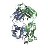 7v64C 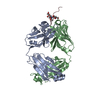 7v7kC 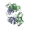 7v8qC 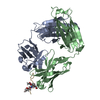 7vacC 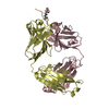 7vazC 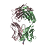 4yhyS S: Starting model for refinement C: citing same article ( |
|---|---|
| Similar structure data |
-Links
 PDBj
PDBj


- Assembly
| Deposited unit | 
| |||||||||||||||||||||||||||||||||||||||||||||||||||||||||||||||||||||||||
|---|---|---|---|---|---|---|---|---|---|---|---|---|---|---|---|---|---|---|---|---|---|---|---|---|---|---|---|---|---|---|---|---|---|---|---|---|---|---|---|---|---|---|---|---|---|---|---|---|---|---|---|---|---|---|---|---|---|---|---|---|---|---|---|---|---|---|---|---|---|---|---|---|---|---|
| 1 | 
| |||||||||||||||||||||||||||||||||||||||||||||||||||||||||||||||||||||||||
| 2 | 
| |||||||||||||||||||||||||||||||||||||||||||||||||||||||||||||||||||||||||
| Unit cell |
| |||||||||||||||||||||||||||||||||||||||||||||||||||||||||||||||||||||||||
| Noncrystallographic symmetry (NCS) | NCS domain:
NCS domain segments:
NCS ensembles :
|
-Components
| #1: Antibody | Mass: 23543.229 Da / Num. of mol.: 2 Source method: isolated from a genetically manipulated source Source: (gene. exp.)  Trichopalpus nigribasis (fry) Trichopalpus nigribasis (fry)#2: Antibody | Mass: 23986.049 Da / Num. of mol.: 2 Source method: isolated from a genetically manipulated source Source: (gene. exp.) Trichopalpus nigribasis (fry)Has protein modification | Y | |
|---|
-Experimental details
-Experiment
| Experiment | Method: X-RAY DIFFRACTION / Number of used crystals: 1 |
|---|
- Sample preparation
| Crystal | Density Matthews: 2.3 Å3/Da / Density % sol: 46.52 % |
|---|---|
| Crystal grow | Temperature: 289 K / Method: vapor diffusion, hanging drop / pH: 8.5 Details: 1.0 M Lithium chloride 0.1 M Tris pH 8.5 20%(w/v) PEG 6000 |
-Data collection
| Diffraction | Mean temperature: 100 K / Serial crystal experiment: N |
|---|---|
| Diffraction source | Source: ROTATING ANODE / Type: RIGAKU / Wavelength: 1.54 Å |
| Detector | Type: RIGAKU RAXIS IV++ / Detector: IMAGE PLATE / Date: Sep 19, 2017 |
| Radiation | Protocol: SINGLE WAVELENGTH / Monochromatic (M) / Laue (L): M / Scattering type: x-ray |
| Radiation wavelength | Wavelength: 1.54 Å / Relative weight: 1 |
| Reflection | Resolution: 2.98→48.68 Å / Num. obs: 18236 / % possible obs: 97.7 % / Redundancy: 4.1 % / Biso Wilson estimate: 42.91 Å2 / CC1/2: 0.97 / Rpim(I) all: 0.086 / Rrim(I) all: 0.184 / Net I/σ(I): 9.1 |
| Reflection shell | Resolution: 2.98→3.09 Å / Redundancy: 3.9 % / Mean I/σ(I) obs: 2.1 / Num. unique obs: 1686 / CC1/2: 0.707 / Rpim(I) all: 0.35 / Rrim(I) all: 0.728 / % possible all: 92.3 |
- Processing
| Software |
| |||||||||||||||||||||||||||||||||||||||||||||||||
|---|---|---|---|---|---|---|---|---|---|---|---|---|---|---|---|---|---|---|---|---|---|---|---|---|---|---|---|---|---|---|---|---|---|---|---|---|---|---|---|---|---|---|---|---|---|---|---|---|---|---|
| Refinement | Method to determine structure: MOLECULAR REPLACEMENT Starting model: 4YHY Resolution: 2.98→48.68 Å / SU ML: 0.2622 / Cross valid method: FREE R-VALUE / σ(F): 1.34 / Phase error: 24.0908 Stereochemistry target values: GeoStd + Monomer Library + CDL v1.2
| |||||||||||||||||||||||||||||||||||||||||||||||||
| Solvent computation | Shrinkage radii: 0.9 Å / VDW probe radii: 1.11 Å / Solvent model: FLAT BULK SOLVENT MODEL | |||||||||||||||||||||||||||||||||||||||||||||||||
| Displacement parameters | Biso mean: 37.69 Å2 | |||||||||||||||||||||||||||||||||||||||||||||||||
| Refinement step | Cycle: LAST / Resolution: 2.98→48.68 Å
| |||||||||||||||||||||||||||||||||||||||||||||||||
| Refine LS restraints |
| |||||||||||||||||||||||||||||||||||||||||||||||||
| LS refinement shell |
| |||||||||||||||||||||||||||||||||||||||||||||||||
| Refinement TLS params. | Method: refined / Origin x: 56.2554536026 Å / Origin y: 36.6470574259 Å / Origin z: 195.849289822 Å
| |||||||||||||||||||||||||||||||||||||||||||||||||
| Refinement TLS group | Selection details: all |
