 Movie
Movie Controller

[English] 日本語
 Yorodumi
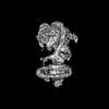
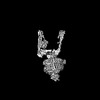
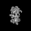
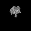
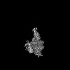
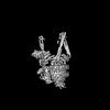
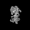
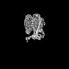
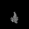
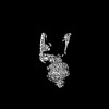
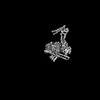
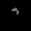
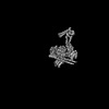
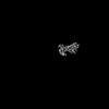
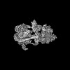
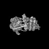
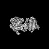
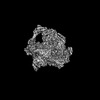
- PDB-7uzk: Rat Kidney V1 complex lacking subunit H with SidK and NCOA7B, State 1 -
+ Open data

- Basic information
| Entry | Database: PDB / ID: 7uzk | ||||||
|---|---|---|---|---|---|---|---|
| Title | Rat Kidney V1 complex lacking subunit H with SidK and NCOA7B, State 1 | ||||||
 Components Components |
| ||||||
 Keywords Keywords | PROTON TRANSPORT / V-ATPase / Complex / Membrane protein | ||||||
| Function / homology |  Function and homology information Function and homology informationIon channel transport / Transferrin endocytosis and recycling / Amino acids regulate mTORC1 / symbiont-mediated suppression of host phagosome acidification / Insulin receptor recycling / proton-transporting V-type ATPase, V1 domain / synaptic vesicle lumen acidification / cellular response to increased oxygen levels / extrinsic component of synaptic vesicle membrane / vacuolar proton-transporting V-type ATPase, V1 domain ...Ion channel transport / Transferrin endocytosis and recycling / Amino acids regulate mTORC1 / symbiont-mediated suppression of host phagosome acidification / Insulin receptor recycling / proton-transporting V-type ATPase, V1 domain / synaptic vesicle lumen acidification / cellular response to increased oxygen levels / extrinsic component of synaptic vesicle membrane / vacuolar proton-transporting V-type ATPase, V1 domain / clathrin-coated vesicle membrane / proton-transporting V-type ATPase complex / protein localization to cilium / vacuolar proton-transporting V-type ATPase complex / vacuolar acidification / : / ROS and RNS production in phagocytes / Neutrophil degranulation / ATPase complex / homeostatic process / microvillus / proton-transporting ATPase activity, rotational mechanism / cilium assembly / H+-transporting two-sector ATPase / ATP metabolic process / ruffle / proton transmembrane transport / apical part of cell / melanosome / ATPase binding / presynapse / intracellular iron ion homeostasis / endosome / apical plasma membrane / cilium / lysosomal membrane / centrosome / ATP hydrolysis activity / nucleoplasm / ATP binding / membrane / plasma membrane / cytoplasm / cytosol Similarity search - Function | ||||||
| Biological species |   Legionella pneumophila (bacteria) Legionella pneumophila (bacteria) | ||||||
| Method | ELECTRON MICROSCOPY / single particle reconstruction / cryo EM / Resolution: 3 Å | ||||||
 Authors Authors | Abbas, Y.M. / Rubinstein, J.L. | ||||||
| Funding support |  Canada, 1items Canada, 1items
| ||||||
 Citation Citation | Journal: To Be Published Title: Structure of V-ATPase with NCOA7B Authors: Abbas, Y.M. / Rubinstein, J.L. | ||||||
| History |
|
- Structure visualization
| Structure viewer | Molecule: MolmilJmol/JSmol |
|---|
- Downloads & links
-Download
| PDBx/mmCIF format | 7uzk.cif.gz | 1.1 MB | Display | PDBx/mmCIF format |
|---|---|---|---|---|
| PDB format | pdb7uzk.ent.gz | Display | PDB format | |
| PDBx/mmJSON format | 7uzk.json.gz | Tree view | PDBx/mmJSON format | |
| Others |  Other downloads Other downloads |
-Validation report
| Arichive directory | https://data.pdbj.org/pub/pdb/validation_reports/uz/7uzkftp://data.pdbj.org/pub/pdb/validation_reports/uz/7uzk | HTTPS FTP |
|---|
-Related structure data
| Related structure data |  26914MC  7uzfC  7uzgC  7uzhC 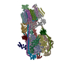 7uziC  7uzjC C: citing same article ( M: map data used to model this data |
|---|---|
| Similar structure data |


-Links
 PDBj
PDBj


- Assembly
| Deposited unit | 
|
|---|---|
| 1 |
|
-Components
-ATPase H+-transporting V1 subunit ... , 2 types, 4 molecules ABCH
| #1: Protein | Mass: 68341.836 Da / Num. of mol.: 3 / Source method: isolated from a natural source / Source: (natural) #4: Protein | | Mass: 28359.020 Da / Num. of mol.: 1 / Source method: isolated from a natural source / Source: (natural) |
|---|
-V-type proton ATPase subunit ... , 5 types, 11 molecules DEFGIJKLMNO
| #2: Protein | Mass: 56611.570 Da / Num. of mol.: 3 / Source method: isolated from a natural source / Source: (natural) #3: Protein | | Mass: 43958.453 Da / Num. of mol.: 1 / Source method: isolated from a natural source / Source: (natural) #5: Protein | Mass: 26167.453 Da / Num. of mol.: 3 / Source method: isolated from a natural source / Source: (natural) #6: Protein | | Mass: 13389.262 Da / Num. of mol.: 1 / Source method: isolated from a natural source / Source: (natural) #7: Protein | Mass: 13733.393 Da / Num. of mol.: 3 / Source method: isolated from a natural source / Source: (natural) |
|---|
-Protein , 2 types, 4 molecules QRST
| #8: Protein | Mass: 32146.178 Da / Num. of mol.: 3 Source method: isolated from a genetically manipulated source Source: (gene. exp.) Legionella pneumophila (bacteria) / Gene: lpg0968 / Production host: #9: Protein | | Mass: 25596.682 Da / Num. of mol.: 1 Source method: isolated from a genetically manipulated source Source: (gene. exp.) |
|---|
-Non-polymers , 2 types, 2 molecules 

| #10: Chemical | ChemComp-ADP /  Mass: 427.201 Da / Num. of mol.: 1 / Source method: obtained synthetically / Formula: C10H15N5O10P2 / Feature type: SUBJECT OF INVESTIGATION / Comment: ADP, energy-carrying molecule*YM Mass: 427.201 Da / Num. of mol.: 1 / Source method: obtained synthetically / Formula: C10H15N5O10P2 / Feature type: SUBJECT OF INVESTIGATION / Comment: ADP, energy-carrying molecule*YM |
|---|---|
| #11: Chemical | ChemComp-MG /  Mass: 24.305 Da / Num. of mol.: 1 / Source method: obtained synthetically / Formula: Mg / Feature type: SUBJECT OF INVESTIGATION Mass: 24.305 Da / Num. of mol.: 1 / Source method: obtained synthetically / Formula: Mg / Feature type: SUBJECT OF INVESTIGATION |
-Details
| Has ligand of interest | Y |
|---|
-Experimental details
-Experiment
| Experiment | Method: ELECTRON MICROSCOPY |
|---|---|
| EM experiment | Aggregation state: PARTICLE / 3D reconstruction method: single particle reconstruction |
- Sample preparation
| Component | Name: Rat Kidney V1 Complex with SidK and NCOA7B / Type: COMPLEX / Entity ID: #1-#9 / Source: MULTIPLE SOURCES |
|---|---|
| Source (natural) | Organism: |
| Buffer solution | pH: 7 |
| Specimen | Embedding applied: NO / Shadowing applied: NO / Staining applied: NO / Vitrification applied: YES |
| Vitrification | Cryogen name: ETHANE |
- Electron microscopy imaging
| Experimental equipment |  Model: Titan Krios / Image courtesy: FEI Company |
|---|---|
| Microscopy | Model: FEI TITAN KRIOS |
| Electron gun | Electron source:  FIELD EMISSION GUN / Accelerating voltage: 300 kV / Illumination mode: FLOOD BEAM FIELD EMISSION GUN / Accelerating voltage: 300 kV / Illumination mode: FLOOD BEAM |
| Electron lens | Mode: BRIGHT FIELD / Nominal defocus max: 3800 nm / Nominal defocus min: 300 nm |
| Image recording | Electron dose: 40.826 e/Å2 / Film or detector model: FEI FALCON IV (4k x 4k) |
- Processing
| CTF correction | Type: PHASE FLIPPING AND AMPLITUDE CORRECTION |
|---|---|
| 3D reconstruction | Resolution: 3 Å / Resolution method: OTHER / Num. of particles: 175138 Details: Model-map FSC (threshold 0.5) calculated in phenix.validation_cryoem Symmetry type: POINT |
