[English] 日本語
 Yorodumi
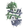
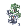
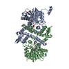
- PDB-7ux7: Crystal structure of MfnG, an L- and D-tyrosine O-methyltransfera... -
+ Open data
- Basic information
| Entry | Database: PDB / ID: 7ux7 | ||||||||||||||||||||||||||||||
|---|---|---|---|---|---|---|---|---|---|---|---|---|---|---|---|---|---|---|---|---|---|---|---|---|---|---|---|---|---|---|---|
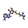
| Title | Crystal structure of MfnG, an L- and D-tyrosine O-methyltransferase from the marformycin biosynthesis pathway of Streptomyces drozdowiczii, with SAH bound at 1.2 A resolution (P212121 - form II) | ||||||||||||||||||||||||||||||
 Components Components | MfnG | ||||||||||||||||||||||||||||||
 Keywords Keywords | TRANSFERASE / O-methyltransferase / O-methyl-tyrosine / Marformycin synthesis / SAM-dependent methyltransferase | ||||||||||||||||||||||||||||||
| Function / homology |  Function and homology information Function and homology informationO-methyltransferase activity / methylation / protein dimerization activity Similarity search - Function | ||||||||||||||||||||||||||||||
| Biological species |  Streptomyces drozdowiczii (bacteria) Streptomyces drozdowiczii (bacteria) | ||||||||||||||||||||||||||||||
| Method |  X-RAY DIFFRACTION / X-RAY DIFFRACTION /  SYNCHROTRON / SYNCHROTRON /  MOLECULAR REPLACEMENT / MOLECULAR REPLACEMENT /  molecular replacement / Resolution: 1.14 Å molecular replacement / Resolution: 1.14 Å | ||||||||||||||||||||||||||||||
 Authors Authors | Miller, M.D. / Wu, K.-L. / Xu, W. / Xiao, H. / Philips Jr., G.N. | ||||||||||||||||||||||||||||||
| Funding support |  United States, 9items United States, 9items
| ||||||||||||||||||||||||||||||
 Citation Citation |  Journal: Protein Sci. / Year: 2022 Journal: Protein Sci. / Year: 2022Title: Expanding the eukaryotic genetic code with a biosynthesized 21st amino acid. Authors: Wu, K.L. / Moore, J.A. / Miller, M.D. / Chen, Y. / Lee, C. / Xu, W. / Peng, Z. / Duan, Q. / Phillips Jr., G.N. / Uribe, R.A. / Xiao, H. | ||||||||||||||||||||||||||||||
| History |
|
- Structure visualization
| Structure viewer | Molecule:  Molmil Molmil Jmol/JSmol Jmol/JSmol |
|---|
- Downloads & links
- Download
| PDBx/mmCIF format |  7ux7.cif.gz 7ux7.cif.gz | 485.9 KB | Display |  PDBx/mmCIF format PDBx/mmCIF format |
|---|---|---|---|---|
| PDB format |  pdb7ux7.ent.gz pdb7ux7.ent.gz | 407.1 KB | Display |  PDB format PDB format |
| PDBx/mmJSON format |  7ux7.json.gz 7ux7.json.gz | Tree view |  PDBx/mmJSON format PDBx/mmJSON format | |
| Others |  Other downloads Other downloads |
-Validation report
| Summary document |  7ux7_validation.pdf.gz 7ux7_validation.pdf.gz | 951.8 KB | Display |  wwPDB validaton report wwPDB validaton report |
|---|---|---|---|---|
| Full document |  7ux7_full_validation.pdf.gz 7ux7_full_validation.pdf.gz | 959.2 KB | Display | |
| Data in XML |  7ux7_validation.xml.gz 7ux7_validation.xml.gz | 36.7 KB | Display | |
| Data in CIF |  7ux7_validation.cif.gz 7ux7_validation.cif.gz | 57.7 KB | Display | |
| Arichive directory |  https://data.pdbj.org/pub/pdb/validation_reports/ux/7ux7 https://data.pdbj.org/pub/pdb/validation_reports/ux/7ux7 ftp://data.pdbj.org/pub/pdb/validation_reports/ux/7ux7 ftp://data.pdbj.org/pub/pdb/validation_reports/ux/7ux7 | HTTPS FTP |
-Related structure data
| Related structure data |  7ux6SC  7ux8C S: Starting model for refinement C: citing same article ( |
|---|---|
| Similar structure data | Similarity search - Function & homology  F&H Search F&H Search |
- Links
Links
- Assembly
| Deposited unit | 
| ||||||||
|---|---|---|---|---|---|---|---|---|---|
| 1 |
| ||||||||
| Unit cell |
|
- Components
| #1: Protein | Mass: 42042.246 Da / Num. of mol.: 2 Source method: isolated from a genetically manipulated source Source: (gene. exp.)  Streptomyces drozdowiczii (bacteria) / Plasmid: pET22b-T5-MfnG-TEV-His / Production host: Streptomyces drozdowiczii (bacteria) / Plasmid: pET22b-T5-MfnG-TEV-His / Production host:  #2: Chemical | #3: Chemical | Num. of mol.: 2 / Source method: obtained synthetically #4: Water | ChemComp-HOH / | Has ligand of interest | Y | Sequence details | The construct was cloned into the EcoRI and HindIII sites of pET22b with an N-terminal Met (0) ...The construct was cloned into the EcoRI and HindIII sites of pET22b with an N-terminal Met (0) added and the native GUG-start codon being expressed as V. A C-terminal expression and 6-His tag ASENLYFQ/GGGHHHHHHG | |
|---|
-Experimental details
-Experiment
| Experiment | Method:  X-RAY DIFFRACTION / Number of used crystals: 1 X-RAY DIFFRACTION / Number of used crystals: 1 |
|---|
- Sample preparation
| Crystal | Density Matthews: 2.1 Å3/Da / Density % sol: 41.3 % |
|---|---|
| Crystal grow | Temperature: 293 K / Method: vapor diffusion, sitting drop / pH: 8 Details: 0.1 M Tris pH 8, 30% Polyethylene glycol monomethyl ether (PEG MME) 2000, Additive: 0.002 M S-Adenosyl methionine (SAM/AdoMet) |
-Data collection
| Diffraction | Mean temperature: 100 K Crystal treatment: flash cooled by immersion in liquid nitrogen Serial crystal experiment: N |
|---|---|
| Diffraction source | Source:  SYNCHROTRON / Site: SYNCHROTRON / Site:  APS APS  / Beamline: 21-ID-D / Wavelength: 0.9762 Å / Beamline: 21-ID-D / Wavelength: 0.9762 Å |
| Detector | Type: DECTRIS EIGER X 9M / Detector: PIXEL / Date: Feb 10, 2021 |
| Radiation | Monochromator: Si(111) / Protocol: SINGLE WAVELENGTH / Monochromatic (M) / Laue (L): M / Scattering type: x-ray |
| Radiation wavelength | Wavelength: 0.9762 Å / Relative weight: 1 |
| Reflection | Resolution: 1.139→72.655 Å / Num. obs: 214260 / % possible obs: 89.6 % / Redundancy: 32.75 % / Biso Wilson estimate: 11.85 Å2 Details: merged and scaled data post-processed by STARANISO for conversion from intensities to structure factor amplitudes CC1/2: 0.996 / Rmerge(I) obs: 0.143 / Rpim(I) all: 0.0248 / Rrim(I) all: 0.1452 / Net I/σ(I): 15.13 / Num. measured all: 7017400 |
| Reflection shell | Resolution: 1.139→1.204 Å / Redundancy: 23.24 % / Rmerge(I) obs: 1.5681 / Mean I/σ(I) obs: 2.17 / Num. measured all: 247895 / Num. measured obs: 247895 / Num. unique all: 10668 / Num. unique obs: 10668 / CC1/2: 0.727 / Rpim(I) all: 0.3222 / Rrim(I) all: 1.6026 / % possible all: 42.5 |
-Phasing
| Phasing | Method:  molecular replacement molecular replacement |
|---|
- Processing
| Software |
| |||||||||||||||||||||||||||||||||||||||||||||||||||||||||||||||||||||||||||||||||||||||||||||||||||||||||||||||||||||||||||||||||||||||||||||||||||||||||||||||||||||||||||||||||||||||||||||||||||||||||||||||||||||||||
|---|---|---|---|---|---|---|---|---|---|---|---|---|---|---|---|---|---|---|---|---|---|---|---|---|---|---|---|---|---|---|---|---|---|---|---|---|---|---|---|---|---|---|---|---|---|---|---|---|---|---|---|---|---|---|---|---|---|---|---|---|---|---|---|---|---|---|---|---|---|---|---|---|---|---|---|---|---|---|---|---|---|---|---|---|---|---|---|---|---|---|---|---|---|---|---|---|---|---|---|---|---|---|---|---|---|---|---|---|---|---|---|---|---|---|---|---|---|---|---|---|---|---|---|---|---|---|---|---|---|---|---|---|---|---|---|---|---|---|---|---|---|---|---|---|---|---|---|---|---|---|---|---|---|---|---|---|---|---|---|---|---|---|---|---|---|---|---|---|---|---|---|---|---|---|---|---|---|---|---|---|---|---|---|---|---|---|---|---|---|---|---|---|---|---|---|---|---|---|---|---|---|---|---|---|---|---|---|---|---|---|---|---|---|---|---|---|---|---|
| Refinement | Method to determine structure:  MOLECULAR REPLACEMENT MOLECULAR REPLACEMENTStarting model: 7UX6 Resolution: 1.14→35.18 Å / SU ML: 0.08 / Cross valid method: THROUGHOUT / σ(F): 1.35 / Phase error: 13.04 / Stereochemistry target values: ML Details: 1. HYDROGENS HAVE BEEN INCLUDED AT THEIR RIDING POSITIONS USING THE ELECTRON CLOUD DISTANCES. 2. THE STRUCTURE CONTAINS AND UNKNOWN LIGAND UNL BOUND IN THE ACTIVE SITE. THE COMPOUND IS A ...Details: 1. HYDROGENS HAVE BEEN INCLUDED AT THEIR RIDING POSITIONS USING THE ELECTRON CLOUD DISTANCES. 2. THE STRUCTURE CONTAINS AND UNKNOWN LIGAND UNL BOUND IN THE ACTIVE SITE. THE COMPOUND IS A METABOLITE THAT CO-PURIFIED WITH THE PROTEIN. THE STRUCTURE LOOKS SIMILAR TO NIACIN, NICOTINAMIDE, BENZOATE, NITORBENZENE ETC. SINCE THE PRECIENSE SPECIES IS NOT KNOW, IT WAS MODELED AS AN UNL. 3. THE C-TERMINUS OF CHAIN A PACKS AGAINST CHAIN B FROM A SYMMETRY MATE IN THE UNIT CELL. THIS SHIFTS THE POSITION OF SEVERAL RESIDUES IN CHAIN B TO ACCOMMODATE THE C-TERMINAL TAG REGION. HOWEVER, THE INTERACTION IS NOT COMPLETE. AFTER MODELING THIS MAJOR SHIFTED PORTION, THERE IS RESIDUAL DIFFERENCE DENSITY FOR A CONFORMATION THAT IS SIMILAR TO THE WHAT IS SEEN IN CHAIN A AND OTHER CRYSTAL FORMS. OCCUPANCY REFINEMENT SUGGESTED ABOUT 60:40 SPLIT. SINCE ONLY THE MAJOR PORTION OF THE CHAIN A C-TERMINUS (367-383)COULD BE MODELED, THIS WAS FIXED AT 0.6 OCCUPANCY TO MATCH THE CHAIN B PORTION THAT IT INTERACTS WITH, WHILE THE OTHER 0.4 OCCUPANCY PORTION COULD NOT BE MODELED DUE TO DISORDER. 4. EVEN THOUGH THE CRYSTALLIZATION DROPS WERE SETUP WITH S-ADENOSYLMETHIONINE (SAM/ ADOMET), ELECTRON DENSITY CLEARLY SHOWS THAT THE COFACTOR HAS BROKEN DOWN TO S-ADENOSYL-L-HOMOCYSTEINE (SAH/ADOHCY). 5. THE DATA WERE PROCESSED WITH STARTANISO DUE TO THE ANISOTROPIC RESOLUTION EXTENT.
| |||||||||||||||||||||||||||||||||||||||||||||||||||||||||||||||||||||||||||||||||||||||||||||||||||||||||||||||||||||||||||||||||||||||||||||||||||||||||||||||||||||||||||||||||||||||||||||||||||||||||||||||||||||||||
| Solvent computation | Shrinkage radii: 0.9 Å / VDW probe radii: 1.11 Å / Solvent model: FLAT BULK SOLVENT MODEL | |||||||||||||||||||||||||||||||||||||||||||||||||||||||||||||||||||||||||||||||||||||||||||||||||||||||||||||||||||||||||||||||||||||||||||||||||||||||||||||||||||||||||||||||||||||||||||||||||||||||||||||||||||||||||
| Displacement parameters | Biso mean: 18.98 Å2 | |||||||||||||||||||||||||||||||||||||||||||||||||||||||||||||||||||||||||||||||||||||||||||||||||||||||||||||||||||||||||||||||||||||||||||||||||||||||||||||||||||||||||||||||||||||||||||||||||||||||||||||||||||||||||
| Refinement step | Cycle: LAST / Resolution: 1.14→35.18 Å
| |||||||||||||||||||||||||||||||||||||||||||||||||||||||||||||||||||||||||||||||||||||||||||||||||||||||||||||||||||||||||||||||||||||||||||||||||||||||||||||||||||||||||||||||||||||||||||||||||||||||||||||||||||||||||
| Refine LS restraints |
| |||||||||||||||||||||||||||||||||||||||||||||||||||||||||||||||||||||||||||||||||||||||||||||||||||||||||||||||||||||||||||||||||||||||||||||||||||||||||||||||||||||||||||||||||||||||||||||||||||||||||||||||||||||||||
| LS refinement shell |
|
 Movie
Movie Controller


 PDBj
PDBj


