 Movie
Movie Controller


+ Open data

- Basic information
| Entry | Database: PDB / ID: 7usf | ||||||||||||
|---|---|---|---|---|---|---|---|---|---|---|---|---|---|
| Title | Mouse mammary tumor virus strand transfer complex intasome | ||||||||||||
 Components Components |
| ||||||||||||
 Keywords Keywords | HYDROLASE/DNA / Integrase-DNA complex / hydrolase / VIRAL PROTEIN / HYDROLASE-DNA complex | ||||||||||||
| Function / homology |  Function and homology information Function and homology informationdUTP diphosphatase activity / DNA integration / viral genome integration into host DNA / establishment of integrated proviral latency / RNA stem-loop binding / RNA-directed DNA polymerase activity / RNA-DNA hybrid ribonuclease activity / viral nucleocapsid / DNA recombination / structural constituent of virion ...dUTP diphosphatase activity / DNA integration / viral genome integration into host DNA / establishment of integrated proviral latency / RNA stem-loop binding / RNA-directed DNA polymerase activity / RNA-DNA hybrid ribonuclease activity / viral nucleocapsid / DNA recombination / structural constituent of virion / aspartic-type endopeptidase activity / DNA-directed DNA polymerase activity / viral translational frameshifting / symbiont entry into host cell / proteolysis / DNA binding / zinc ion binding Similarity search - Function | ||||||||||||
| Biological species |  Mouse mammary tumor virus Mouse mammary tumor virus | ||||||||||||
| Method | ELECTRON MICROSCOPY / single particle reconstruction / cryo EM / Resolution: 3.5 Å | ||||||||||||
 Authors Authors | Jozwik, I. / Lyumkis, D. | ||||||||||||
| Funding support |  United States, 3items United States, 3items
| ||||||||||||
 Citation Citation | Journal: Nucleic Acids Res / Year: 2022 Title: B-to-A transition in target DNA during retroviral integration. Authors: Ilona K Jóźwik / Wen Li / Da-Wei Zhang / Doris Wong / Julia Grawenhoff / Allison Ballandras-Colas / Sriram Aiyer / Peter Cherepanov / Alan N Engelman / Dmitry Lyumkis /   Abstract: Integration into host target DNA (tDNA), a hallmark of retroviral replication, is mediated by the intasome, a multimer of integrase (IN) assembled on viral DNA (vDNA) ends. To ascertain aspects of ...Integration into host target DNA (tDNA), a hallmark of retroviral replication, is mediated by the intasome, a multimer of integrase (IN) assembled on viral DNA (vDNA) ends. To ascertain aspects of tDNA recognition during integration, we have solved the 3.5 Å resolution cryo-EM structure of the mouse mammary tumor virus (MMTV) strand transfer complex (STC) intasome. The tDNA adopts an A-like conformation in the region encompassing the sites of vDNA joining, which exposes the sugar-phosphate backbone for IN-mediated strand transfer. Examination of existing retroviral STC structures revealed conservation of A-form tDNA in the analogous regions of these complexes. Furthermore, analyses of sequence preferences in genomic integration sites selectively targeted by six different retroviruses highlighted consistent propensity for A-philic sequences at the sites of vDNA joining. Our structure additionally revealed several novel MMTV IN-DNA interactions, as well as contacts seen in prior STC structures, including conserved Pro125 and Tyr149 residues interacting with tDNA. In infected cells, Pro125 substitutions impacted the global pattern of MMTV integration without significantly altering local base sequence preferences at vDNA insertion sites. Collectively, these data advance our understanding of retroviral intasome structure and function, as well as factors that influence patterns of vDNA integration in genomic DNA. | ||||||||||||
| History |
|
- Structure visualization
| Structure viewer | Molecule: MolmilJmol/JSmol |
|---|
- Downloads & links
-Download
| PDBx/mmCIF format | 7usf.cif.gz | 224.2 KB | Display | PDBx/mmCIF format |
|---|---|---|---|---|
| PDB format | pdb7usf.ent.gz | 174.2 KB | Display | PDB format |
| PDBx/mmJSON format | 7usf.json.gz | Tree view | PDBx/mmJSON format | |
| Others |  Other downloads Other downloads |
-Validation report
| Arichive directory | https://data.pdbj.org/pub/pdb/validation_reports/us/7usfftp://data.pdbj.org/pub/pdb/validation_reports/us/7usf | HTTPS FTP |
|---|
-Related structure data
| Related structure data |  26737MC  7ut1C M: map data used to model this data C: citing same article ( |
|---|---|
| Similar structure data |
-Links
 PDBj
PDBj


- Assembly
| Deposited unit | 
|
|---|---|
| 1 | 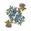
|
| 2 |
|
| 3 | 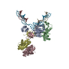
|
| Symmetry | Point symmetry: (Schoenflies symbol: C2 (2 fold cyclic)) |
-Components
-Protein , 1 types, 4 molecules ABCD
| #1: Protein | Mass: 35753.332 Da / Num. of mol.: 4 / Mutation: T252S Source method: isolated from a genetically manipulated source Source: (gene. exp.) Mouse mammary tumor virus / Gene: gag-pro-pol / Production host:  |
|---|
-DNA chain , 3 types, 3 molecules IJK
| #2: DNA chain | Mass: 9234.931 Da / Num. of mol.: 1 / Source method: obtained synthetically / Source: (synth.) Mouse mammary tumor virus |
|---|---|
| #3: DNA chain | Mass: 12875.227 Da / Num. of mol.: 1 / Source method: obtained synthetically / Source: (synth.) Mouse mammary tumor virus |
| #4: DNA chain | Mass: 4980.269 Da / Num. of mol.: 1 / Source method: obtained synthetically / Source: (synth.) Mouse mammary tumor virus |
-Non-polymers , 2 types, 5 molecules 

| #5: Chemical | ChemComp-ZN /  Mass: 65.409 Da / Num. of mol.: 4 / Source method: obtained synthetically / Formula: Zn Mass: 65.409 Da / Num. of mol.: 4 / Source method: obtained synthetically / Formula: Zn#6: Chemical | ChemComp-CA / |  Mass: 40.078 Da / Num. of mol.: 1 / Source method: obtained synthetically / Formula: Ca Mass: 40.078 Da / Num. of mol.: 1 / Source method: obtained synthetically / Formula: Ca |
|---|
-Details
| Has ligand of interest | N |
|---|
-Experimental details
-Experiment
| Experiment | Method: ELECTRON MICROSCOPY |
|---|---|
| EM experiment | Aggregation state: PARTICLE / 3D reconstruction method: single particle reconstruction |
- Sample preparation
| Component | Name: MMTV strand transfer complex intasome / Type: COMPLEX / Entity ID: #1-#4 / Source: RECOMBINANT | ||||||||||||||||||||||||||||||
|---|---|---|---|---|---|---|---|---|---|---|---|---|---|---|---|---|---|---|---|---|---|---|---|---|---|---|---|---|---|---|---|
| Molecular weight | Value: 0.3 kDa/nm / Experimental value: NO | ||||||||||||||||||||||||||||||
| Source (natural) | Organism: Mouse mammary tumor virus | ||||||||||||||||||||||||||||||
| Source (recombinant) | Organism: | ||||||||||||||||||||||||||||||
| Buffer solution | pH: 6.5 | ||||||||||||||||||||||||||||||
| Buffer component |
| ||||||||||||||||||||||||||||||
| Specimen | Embedding applied: NO / Shadowing applied: NO / Staining applied: NO / Vitrification applied: YES Details: MMTV STC intasomes were applied onto R1.2/1.3 gold UltrAufoil grids, Au 300 mesh (Quantifoil). Cryo-EM grids were prepared by manually freezing using a manual plunger in cold room at 4C and ...Details: MMTV STC intasomes were applied onto R1.2/1.3 gold UltrAufoil grids, Au 300 mesh (Quantifoil). Cryo-EM grids were prepared by manually freezing using a manual plunger in cold room at 4C and stored in liquid nitrogen for future data acquisition. | ||||||||||||||||||||||||||||||
| Specimen support | Grid material: GOLD / Grid type: UltrAuFoil | ||||||||||||||||||||||||||||||
| Vitrification | Instrument: HOMEMADE PLUNGER / Cryogen name: ETHANE Details: Cryo-EM grids were prepared by freezing using a manual plunger in cold room at 4C |
- Electron microscopy imaging
| Experimental equipment |  Model: Titan Krios / Image courtesy: FEI Company |
|---|---|
| Microscopy | Model: FEI TITAN KRIOS |
| Electron gun | Electron source:  FIELD EMISSION GUN / Accelerating voltage: 300 kV / Illumination mode: FLOOD BEAM FIELD EMISSION GUN / Accelerating voltage: 300 kV / Illumination mode: FLOOD BEAM |
| Electron lens | Mode: BRIGHT FIELD / Nominal magnification: 22500 X / Calibrated magnification: 38167 X / Nominal defocus max: 3000 nm / Nominal defocus min: 1000 nm / Cs: 2.7 mm / C2 aperture diameter: 70 µm / Alignment procedure: COMA FREE |
| Specimen holder | Cryogen: NITROGEN / Specimen holder model: FEI TITAN KRIOS AUTOGRID HOLDER |
| Image recording | Average exposure time: 10 sec. / Electron dose: 67 e/Å2 / Detector mode: COUNTING / Film or detector model: GATAN K2 SUMMIT (4k x 4k) / Num. of grids imaged: 1 / Num. of real images: 1578 |
| Image scans | Width: 3838 / Height: 3710 / Movie frames/image: 100 / Used frames/image: 1-100 |
- Processing
| EM software |
| ||||||||||||||||||||||||||||||||
|---|---|---|---|---|---|---|---|---|---|---|---|---|---|---|---|---|---|---|---|---|---|---|---|---|---|---|---|---|---|---|---|---|---|
| CTF correction | Type: PHASE FLIPPING AND AMPLITUDE CORRECTION | ||||||||||||||||||||||||||||||||
| Particle selection | Num. of particles selected: 1048508 | ||||||||||||||||||||||||||||||||
| 3D reconstruction | Resolution: 3.5 Å / Resolution method: FSC 0.143 CUT-OFF / Num. of particles: 50196 / Algorithm: FOURIER SPACE / Symmetry type: POINT | ||||||||||||||||||||||||||||||||
| Atomic model building | Protocol: FLEXIBLE FIT / Space: REAL / Target criteria: CC Details: Initial model building was accomplished by rigid-body fitting of the MMTV CSC structure downloaded from the Protein Data Bank (PDB ID: 3JCA) into the EM map in Chimera 1.14 by Fit in Map ...Details: Initial model building was accomplished by rigid-body fitting of the MMTV CSC structure downloaded from the Protein Data Bank (PDB ID: 3JCA) into the EM map in Chimera 1.14 by Fit in Map tool. Unmodeled protein and DNA residues were manually built in Coot 0.9.4.1 and the structure underwent a few iterative cycles of manual model re-building and real-space refinement in Phenix. Ramachandran and secondary structure restraints were applied. To model the full octameric intasome, we additionally rigid-body docked the flanking IN dimers (PDB ID: 5CZ2) into the map. The density connecting the flanking dimers and the core was evident, but broken, and therefore a model was not derived for the linker regions. The final model accounts for the complete octameric MMTV STC intasome with connections for the linker regions. |
