National Institutes of Health/National Institute Of Allergy and Infectious Diseases (NIH/NIAID)
U54 AI150472
United States
National Institutes of Health/National Institute Of Allergy and Infectious Diseases (NIH/NIAID)
R01 AI136680
United States
Citation
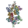
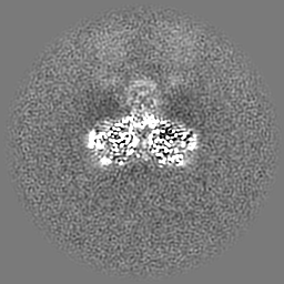
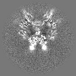
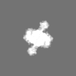
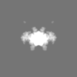
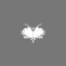
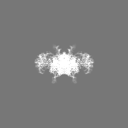
Journal: Nucleic Acids Res / Year: 2022 Title: B-to-A transition in target DNA during retroviral integration. Authors: Ilona K Jóźwik / Wen Li / Da-Wei Zhang / Doris Wong / Julia Grawenhoff / Allison Ballandras-Colas / Sriram Aiyer / Peter Cherepanov / Alan N Engelman / Dmitry Lyumkis / Abstract: Integration into host target DNA (tDNA), a hallmark of retroviral replication, is mediated by the intasome, a multimer of integrase (IN) assembled on viral DNA (vDNA) ends. To ascertain aspects of ...Integration into host target DNA (tDNA), a hallmark of retroviral replication, is mediated by the intasome, a multimer of integrase (IN) assembled on viral DNA (vDNA) ends. To ascertain aspects of tDNA recognition during integration, we have solved the 3.5 Å resolution cryo-EM structure of the mouse mammary tumor virus (MMTV) strand transfer complex (STC) intasome. The tDNA adopts an A-like conformation in the region encompassing the sites of vDNA joining, which exposes the sugar-phosphate backbone for IN-mediated strand transfer. Examination of existing retroviral STC structures revealed conservation of A-form tDNA in the analogous regions of these complexes. Furthermore, analyses of sequence preferences in genomic integration sites selectively targeted by six different retroviruses highlighted consistent propensity for A-philic sequences at the sites of vDNA joining. Our structure additionally revealed several novel MMTV IN-DNA interactions, as well as contacts seen in prior STC structures, including conserved Pro125 and Tyr149 residues interacting with tDNA. In infected cells, Pro125 substitutions impacted the global pattern of MMTV integration without significantly altering local base sequence preferences at vDNA insertion sites. Collectively, these data advance our understanding of retroviral intasome structure and function, as well as factors that influence patterns of vDNA integration in genomic DNA.
Name: ZINC ION / type: ligand / ID: 5 / Number of copies: 4 / Formula: ZN
Molecular weight
Theoretical: 65.409 Da
-
Macromolecule #6: CALCIUM ION
Macromolecule
Name: CALCIUM ION / type: ligand / ID: 6 / Number of copies: 1 / Formula: CA
Molecular weight
Theoretical: 40.078 Da
-
Experimental details
-
Structure determination
Method
cryo EM
Processing
single particle reconstruction
Aggregation state
particle
-
Sample preparation
Buffer
pH: 6.5 Component:
Concentration
Name
Formula
25.0 mM
Tris-HCl
200.0 mM
sodium chloride
NaCl
2.0 mM
DTT
10.0 mM
Calcium chloride
CaCl2
25.0 mM
Zinc chloride
ZnCl2
Grid
Model: UltrAuFoil / Material: GOLD / Support film - Material: GOLD / Support film - topology: HOLEY / Pretreatment - Type: PLASMA CLEANING / Pretreatment - Time: 7 sec.
Vitrification
Cryogen name: ETHANE / Instrument: HOMEMADE PLUNGER Details: Cryo-EM grids were prepared by freezing using a manual plunger in cold room at 4C.
Details
MMTV STC intasomes were applied onto R1.2/1.3 gold UltrAufoil grids, Au 300 mesh (Quantifoil). Cryo-EM grids were prepared by manually freezing using a manual plunger in cold room at 4C and stored in liquid nitrogen for future data acquisition.
-
Electron microscopy
Microscope
FEI TITAN KRIOS
Image recording
Film or detector model: GATAN K2 SUMMIT (4k x 4k) / Detector mode: COUNTING / Digitization - Dimensions - Width: 3838 pixel / Digitization - Dimensions - Height: 3710 pixel / Digitization - Frames/image: 1-100 / Number grids imaged: 1 / Number real images: 1578 / Average exposure time: 10.0 sec. / Average electron dose: 67.0 e/Å2
Electron beam
Acceleration voltage: 300 kV / Electron source: FIELD EMISSION GUN
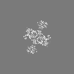
Initial model building was accomplished by rigid-body fitting of the MMTV CSC structure downloaded from the Protein Data Bank (PDB ID: 3JCA) into the EM map in Chimera 1.14 by Fit in Map tool. Unmodeled protein and DNA residues were manually built in Coot 0.9.4.1 and the structure underwent a few iterative cycles of manual model re-building and real-space refinement in Phenix. Ramachandran and secondary structure restraints were applied. To model the full octameric intasome, we additionally rigid-body docked the flanking IN dimers (PDB ID: 5CZ2) into the map. The density connecting the flanking dimers and the core was evident, but broken, and therefore a model was not derived for the linker regions. The final model accounts for the complete octameric MMTV STC intasome with connections for the linker regions.
Refinement
Space: REAL / Protocol: FLEXIBLE FIT / Target criteria: CC
Output model
PDB-7usf: Mouse mammary tumor virus strand transfer complex intasome
+
About Yorodumi
-
News
-
Feb 9, 2022. New format data for meta-information of EMDB entries
New format data for meta-information of EMDB entries
Version 3 of the EMDB header file is now the official format.
The previous official version 1.9 will be removed from the archive.
In the structure databanks used in Yorodumi, some data are registered as the other names, "COVID-19 virus" and "2019-nCoV". Here are the details of the virus and the list of structure data.
Jan 31, 2019. EMDB accession codes are about to change! (news from PDBe EMDB page)
EMDB accession codes are about to change! (news from PDBe EMDB page)
The allocation of 4 digits for EMDB accession codes will soon come to an end. Whilst these codes will remain in use, new EMDB accession codes will include an additional digit and will expand incrementally as the available range of codes is exhausted. The current 4-digit format prefixed with “EMD-” (i.e. EMD-XXXX) will advance to a 5-digit format (i.e. EMD-XXXXX), and so on. It is currently estimated that the 4-digit codes will be depleted around Spring 2019, at which point the 5-digit format will come into force.
The EM Navigator/Yorodumi systems omit the EMD- prefix.
Related info.:Q: What is EMD? / ID/Accession-code notation in Yorodumi/EM Navigator
Yorodumi is a browser for structure data from EMDB, PDB, SASBDB, etc.
This page is also the successor to EM Navigator detail page, and also detail information page/front-end page for Omokage search.
The word "yorodu" (or yorozu) is an old Japanese word meaning "ten thousand". "mi" (miru) is to see.
Related info.:EMDB / PDB / SASBDB / Comparison of 3 databanks / Yorodumi Search / Aug 31, 2016. New EM Navigator & Yorodumi / Yorodumi Papers / Jmol/JSmol / Function and homology information / Changes in new EM Navigator and Yorodumi
 Movie
Movie Controller


 Open data
 Basic information
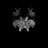
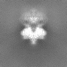
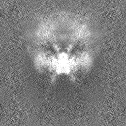
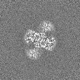
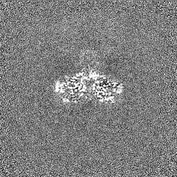
 Map data
 Sample
 Keywords
 Function and homology information
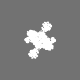
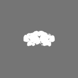
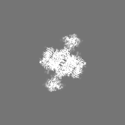
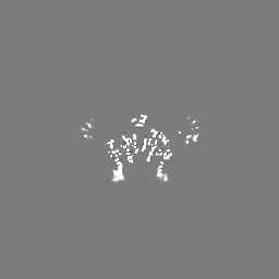
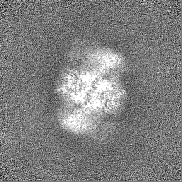
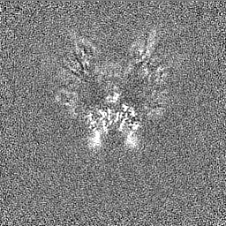
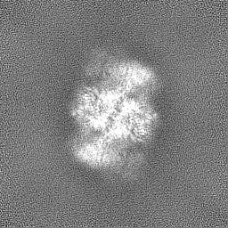
 Mouse mammary tumor virus
 Authors
 United States, 3 items
  Citation

 Structure visualization
 Downloads & links
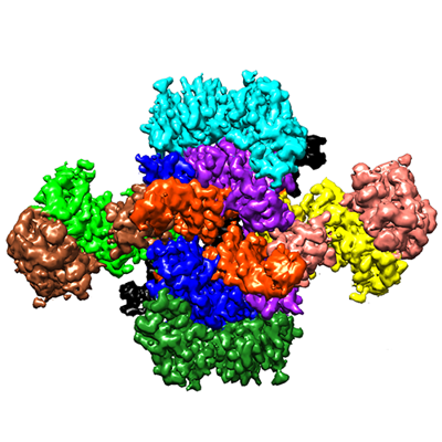 emd_26737.png
 http://ftp.pdbj.org/pub/emdb/structures/EMD-26737


 Z (Sec.)
 Y (Row.)
 X (Col.)


 Sample components
 Processing
 Electron microscopy
 FIELD EMISSION GUN
