[English] 日本語
 Yorodumi
- PDB-7ur4: Cryo-EM Structure of the Neutralizing Antibody MPV467 in Complex ... -
+ Open data
- Basic information
| Entry | Database: PDB / ID: 7ur4 | |||||||||
|---|---|---|---|---|---|---|---|---|---|---|
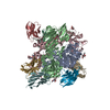
| Title | Cryo-EM Structure of the Neutralizing Antibody MPV467 in Complex with Prefusion Human Metapneumovirus F Glycoprotein | |||||||||
 Components Components |
| |||||||||
 Keywords Keywords | VIRAL PROTEIN/IMMUNE SYSTEM / neutralizing antibody / fusion protein / metapneumovirus / VIRAL PROTEIN-IMMUNE SYSTEM complex | |||||||||
| Function / homology |  Function and homology information Function and homology informationfusion of virus membrane with host plasma membrane / viral envelope / symbiont entry into host cell / host cell plasma membrane / virion membrane / membrane Similarity search - Function | |||||||||
| Biological species |  Human metapneumovirus Human metapneumovirus Homo sapiens (human) Homo sapiens (human) | |||||||||
| Method | ELECTRON MICROSCOPY / single particle reconstruction / cryo EM / Resolution: 3.34 Å | |||||||||
 Authors Authors | Rush, S.A. / McLellan, J.S. | |||||||||
| Funding support |  United States, 2items United States, 2items
| |||||||||
 Citation Citation |  Journal: Proc Natl Acad Sci U S A / Year: 2022 Journal: Proc Natl Acad Sci U S A / Year: 2022Title: Structural basis for ultrapotent antibody-mediated neutralization of human metapneumovirus. Authors: Avik Banerjee / Jiachen Huang / Scott A Rush / Jackelyn Murray / Aaron D Gingerich / Fredejah Royer / Ching-Lin Hsieh / Ralph A Tripp / Jason S McLellan / Jarrod J Mousa /  Abstract: Human metapneumovirus (hMPV) is a leading cause of morbidity and hospitalization among children worldwide, however, no vaccines or therapeutics are currently available for hMPV disease prevention and ...Human metapneumovirus (hMPV) is a leading cause of morbidity and hospitalization among children worldwide, however, no vaccines or therapeutics are currently available for hMPV disease prevention and treatment. The hMPV fusion (F) protein is the sole target of neutralizing antibodies. To map the immunodominant epitopes on the hMPV F protein, we isolated a panel of human monoclonal antibodies (mAbs), and the mAbs were assessed for binding avidity, neutralization potency, and epitope specificity. We found the majority of the mAbs target diverse epitopes on the hMPV F protein, and we discovered multiple mAb binding approaches for antigenic site III. The most potent mAb, MPV467, which had picomolar potency, was examined in prophylactic and therapeutic mouse challenge studies, and MPV467 limited virus replication in mouse lungs when administered 24 h before or 72 h after viral infection. We determined the structure of MPV467 in complex with the hMPV F protein using cryo-electron microscopy to a resolution of 3.3 Å, which revealed a complex novel prefusion-specific epitope overlapping antigenic sites II and V on a single protomer. Overall, our data reveal insights into the immunodominant antigenic epitopes on the hMPV F protein, identify a mAb therapy for hMPV F disease prevention and treatment, and provide the discovery of a prefusion-specific epitope on the hMPV F protein. | |||||||||
| History |
|
- Structure visualization
| Structure viewer | Molecule:  Molmil Molmil Jmol/JSmol Jmol/JSmol |
|---|
- Downloads & links
- Download
| PDBx/mmCIF format |  7ur4.cif.gz 7ur4.cif.gz | 747.7 KB | Display |  PDBx/mmCIF format PDBx/mmCIF format |
|---|---|---|---|---|
| PDB format |  pdb7ur4.ent.gz pdb7ur4.ent.gz | 610.2 KB | Display |  PDB format PDB format |
| PDBx/mmJSON format |  7ur4.json.gz 7ur4.json.gz | Tree view |  PDBx/mmJSON format PDBx/mmJSON format | |
| Others |  Other downloads Other downloads |
-Validation report
| Summary document |  7ur4_validation.pdf.gz 7ur4_validation.pdf.gz | 1.4 MB | Display |  wwPDB validaton report wwPDB validaton report |
|---|---|---|---|---|
| Full document |  7ur4_full_validation.pdf.gz 7ur4_full_validation.pdf.gz | 1.4 MB | Display | |
| Data in XML |  7ur4_validation.xml.gz 7ur4_validation.xml.gz | 63.4 KB | Display | |
| Data in CIF |  7ur4_validation.cif.gz 7ur4_validation.cif.gz | 96.9 KB | Display | |
| Arichive directory |  https://data.pdbj.org/pub/pdb/validation_reports/ur/7ur4 https://data.pdbj.org/pub/pdb/validation_reports/ur/7ur4 ftp://data.pdbj.org/pub/pdb/validation_reports/ur/7ur4 ftp://data.pdbj.org/pub/pdb/validation_reports/ur/7ur4 | HTTPS FTP |
-Related structure data
| Related structure data |  26704MC M: map data used to model this data C: citing same article ( |
|---|---|
| Similar structure data | Similarity search - Function & homology  F&H Search F&H Search |
- Links
Links
- Assembly
| Deposited unit | 
|
|---|---|
| 1 |
|
- Components
| #1: Protein | Mass: 60489.930 Da / Num. of mol.: 3 Mutation: Q100R, S101R, A185P, G294E, A140C, A147C, L110C, N322C, T127C, N153C, T365C, V463C, L219K, V231I, E453Q, V84C, A249C Source method: isolated from a genetically manipulated source Source: (gene. exp.)  Human metapneumovirus / Production host: Human metapneumovirus / Production host:  Homo sapiens (human) / References: UniProt: H6X1Z1 Homo sapiens (human) / References: UniProt: H6X1Z1#2: Antibody | Mass: 24023.779 Da / Num. of mol.: 3 Source method: isolated from a genetically manipulated source Source: (gene. exp.)  Homo sapiens (human) / Production host: Homo sapiens (human) / Production host:  Homo sapiens (human) Homo sapiens (human)#3: Antibody | Mass: 22621.924 Da / Num. of mol.: 3 Source method: isolated from a genetically manipulated source Source: (gene. exp.)  Homo sapiens (human) / Production host: Homo sapiens (human) / Production host:  Homo sapiens (human) Homo sapiens (human)#4: Polysaccharide | Source method: isolated from a genetically manipulated source #5: Sugar | ChemComp-NAG / Has ligand of interest | N | Has protein modification | Y | |
|---|
-Experimental details
-Experiment
| Experiment | Method: ELECTRON MICROSCOPY |
|---|---|
| EM experiment | Aggregation state: PARTICLE / 3D reconstruction method: single particle reconstruction |
- Sample preparation
| Component | Name: Trimeric prefusion hMPV F glycoprotein bound by three molecules of MPV467 Fab Type: COMPLEX / Entity ID: #1-#3 / Source: MULTIPLE SOURCES | |||||||||||||||||||||||||
|---|---|---|---|---|---|---|---|---|---|---|---|---|---|---|---|---|---|---|---|---|---|---|---|---|---|---|
| Source (natural) | Organism:  Human metapneumovirus / Strain: NL/1/00 Human metapneumovirus / Strain: NL/1/00 | |||||||||||||||||||||||||
| Source (recombinant) | Organism:  Homo sapiens (human) Homo sapiens (human) | |||||||||||||||||||||||||
| Buffer solution | pH: 8 | |||||||||||||||||||||||||
| Buffer component |
| |||||||||||||||||||||||||
| Specimen | Embedding applied: NO / Shadowing applied: NO / Staining applied: NO / Vitrification applied: YES | |||||||||||||||||||||||||
| Specimen support | Grid material: GOLD / Grid mesh size: 300 divisions/in. / Grid type: C-flat-1.2/1.3 | |||||||||||||||||||||||||
| Vitrification | Instrument: FEI VITROBOT MARK IV / Cryogen name: ETHANE / Humidity: 100 % / Chamber temperature: 283.15 K |
- Electron microscopy imaging
| Microscopy | Model: TFS GLACIOS |
|---|---|
| Electron gun | Electron source:  FIELD EMISSION GUN / Accelerating voltage: 200 kV / Illumination mode: FLOOD BEAM FIELD EMISSION GUN / Accelerating voltage: 200 kV / Illumination mode: FLOOD BEAM |
| Electron lens | Mode: BRIGHT FIELD / Nominal magnification: 150000 X / Nominal defocus max: 2500 nm / Nominal defocus min: 1000 nm / Cs: 2.7 mm |
| Specimen holder | Cryogen: NITROGEN |
| Image recording | Electron dose: 40 e/Å2 / Film or detector model: FEI FALCON IV (4k x 4k) |
- Processing
| EM software |
| |||||||||||||||
|---|---|---|---|---|---|---|---|---|---|---|---|---|---|---|---|---|
| CTF correction | Type: PHASE FLIPPING AND AMPLITUDE CORRECTION | |||||||||||||||
| Particle selection | Num. of particles selected: 807472 | |||||||||||||||
| 3D reconstruction | Resolution: 3.34 Å / Resolution method: FSC 0.143 CUT-OFF / Num. of particles: 85671 / Symmetry type: POINT |
 Movie
Movie Controller


 PDBj
PDBj


