Entry Database : PDB / ID : 7um3Title Crystal structure of a Fab in complex with a peptide derived from the LAG-3 D1 domain loop insertion D1 domain loop peptide from Lymphocyte activation gene 3 protein Fab heavy chain Fab light chain Keywords / / / / Function / homology Function Domain/homology Component
/ / / / / / / / / / / / / / / / / / / / / / / / / / / / / / / / / / / / Biological species Homo sapiens (human)Method / / / Resolution : 2.3983 Å Authors Zorn, J.A. / Lee, P.S. / Rajpal, A. / Strop, P. Funding support 1items Organization Grant number Country Other private
Journal : Cancer Immunol Res / Year : 2022Title : Preclinical Characterization of Relatlimab, a Human LAG-3-Blocking Antibody, Alone or in Combination with Nivolumab.Authors : Thudium, K. / Selby, M. / Zorn, J.A. / Rak, G. / Wang, X.T. / Bunch, R.T. / Hogan, J.M. / Strop, P. / Korman, A.J. History Deposition Apr 6, 2022 Deposition site / Processing site Revision 1.0 Sep 7, 2022 Provider / Type Revision 1.1 Oct 12, 2022 Group / Category Item / _citation.page_first / _citation.page_lastRevision 1.2 Oct 18, 2023 Group / Refinement descriptionCategory chem_comp_atom / chem_comp_bond ... chem_comp_atom / chem_comp_bond / pdbx_initial_refinement_model / struct_ncs_dom_lim Item _struct_ncs_dom_lim.beg_auth_comp_id / _struct_ncs_dom_lim.beg_label_asym_id ... _struct_ncs_dom_lim.beg_auth_comp_id / _struct_ncs_dom_lim.beg_label_asym_id / _struct_ncs_dom_lim.beg_label_comp_id / _struct_ncs_dom_lim.beg_label_seq_id / _struct_ncs_dom_lim.end_auth_comp_id / _struct_ncs_dom_lim.end_label_asym_id / _struct_ncs_dom_lim.end_label_comp_id / _struct_ncs_dom_lim.end_label_seq_id Revision 1.3 Nov 6, 2024 Group / Category / pdbx_modification_feature
Show all Show less
 Movie
Movie Controller

 Yorodumi
 Open data
 Basic information
 Components
 Keywords
 Function and homology information
 Homo sapiens (human)
 X-RAY DIFFRACTION /
  Authors
 Citation
 Structure visualization
 Downloads & links
 Other downloads
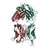
 PDBj
PDBj


 Assembly

