 Movie
Movie Controller

[English] 日本語
 Yorodumi
- PDB-7ulf: l-glutamate/GTP complex of F420-gamma glutamyl ligase (CofE) from... -
+ Open data

- Basic information
| Entry | Database: PDB / ID: 7ulf | ||||||
|---|---|---|---|---|---|---|---|
| Title | l-glutamate/GTP complex of F420-gamma glutamyl ligase (CofE) from Archaeoglobus fulgidus | ||||||
 Components Components | Coenzyme F420:L-glutamate ligase | ||||||
 Keywords Keywords | LIGASE / ligase substrate complex | ||||||
| Function / homology |  Function and homology information Function and homology informationcoenzyme F420-0:L-glutamate ligase / coenzyme F420-1:gamma-L-glutamate ligase / coenzyme F420-0:L-glutamate ligase activity / coenzyme F420-1:gamma-L-glutamate ligase activity / F420-0 metabolic process / GTP binding / metal ion binding Similarity search - Function | ||||||
| Biological species |   Archaeoglobus fulgidus DSM 4304 (archaea) Archaeoglobus fulgidus DSM 4304 (archaea) | ||||||
| Method |  X-RAY DIFFRACTION / SYNCHROTRON / MOLECULAR REPLACEMENT / Resolution: 1.61 Å X-RAY DIFFRACTION / SYNCHROTRON / MOLECULAR REPLACEMENT / Resolution: 1.61 Å | ||||||
 Authors Authors | Bashiri, G. / Squire, C.J. | ||||||
| Funding support | 1items
| ||||||
 Citation Citation | Journal: Nat Commun / Year: 2024 Title: Poly-gamma-glutamylation of biomolecules. Authors: Bashiri, G. / Bulloch, E.M.M. / Bramley, W.R. / Davidson, M. / Stuteley, S.M. / Young, P.G. / Harris, P.W.R. / Naqvi, M.S.H. / Middleditch, M.J. / Schmitz, M. / Chang, W.C. / Baker, E.N. / Squire, C.J. | ||||||
| History |
|
- Structure visualization
| Structure viewer | Molecule: MolmilJmol/JSmol |
|---|
- Downloads & links
-Download
| PDBx/mmCIF format | 7ulf.cif.gz | 67.5 KB | Display | PDBx/mmCIF format |
|---|---|---|---|---|
| PDB format | pdb7ulf.ent.gz | 46.9 KB | Display | PDB format |
| PDBx/mmJSON format | 7ulf.json.gz | Tree view | PDBx/mmJSON format | |
| Others |  Other downloads Other downloads |
-Validation report
| Summary document | 7ulf_validation.pdf.gz | 756.6 KB | Display | wwPDB validaton report |
|---|---|---|---|---|
| Full document | 7ulf_full_validation.pdf.gz | 756.6 KB | Display | |
| Data in XML | 7ulf_validation.xml.gz | 11.6 KB | Display | |
| Data in CIF | 7ulf_validation.cif.gz | 15.9 KB | Display | |
| Arichive directory | https://data.pdbj.org/pub/pdb/validation_reports/ul/7ulfftp://data.pdbj.org/pub/pdb/validation_reports/ul/7ulf | HTTPS FTP |
-Related structure data
| Related structure data |  7uldC 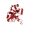 7uleC  8g8pC  2phnS C: citing same article ( S: Starting model for refinement |
|---|---|
| Similar structure data |
-Links
 PDBj
PDBj- Assembly
| Deposited unit | 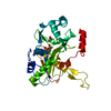
| |||||||||
|---|---|---|---|---|---|---|---|---|---|---|
| 1 | 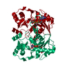
| |||||||||
| Unit cell |
| |||||||||
| Components on special symmetry positions |
|
-Components
| #1: Protein | Mass: 27423.568 Da / Num. of mol.: 1 Source method: isolated from a genetically manipulated source Source: (gene. exp.) Archaeoglobus fulgidus DSM 4304 (archaea)Gene: cofE, AF_2256 / Production host:  References: UniProt: O28028, coenzyme F420-0:L-glutamate ligase, coenzyme F420-1:gamma-L-glutamate ligase | ||||||
|---|---|---|---|---|---|---|---|
| #2: Chemical | ChemComp-GGL /   Type: L-gamma-peptide, C-delta linking / Mass: 147.129 Da / Num. of mol.: 1 / Source method: obtained synthetically / Formula: C5H9NO4 Type: L-gamma-peptide, C-delta linking / Mass: 147.129 Da / Num. of mol.: 1 / Source method: obtained synthetically / Formula: C5H9NO4 | ||||||
| #3: Chemical | ChemComp-GTP /   Mass: 523.180 Da / Num. of mol.: 1 / Source method: obtained synthetically / Formula: C10H16N5O14P3 / Feature type: SUBJECT OF INVESTIGATION / Comment: GTP, energy-carrying molecule*YM Mass: 523.180 Da / Num. of mol.: 1 / Source method: obtained synthetically / Formula: C10H16N5O14P3 / Feature type: SUBJECT OF INVESTIGATION / Comment: GTP, energy-carrying molecule*YM | ||||||
| #4: Chemical |   Mass: 54.938 Da / Num. of mol.: 2 / Source method: obtained synthetically / Formula: Mn Mass: 54.938 Da / Num. of mol.: 2 / Source method: obtained synthetically / Formula: Mn#5: Water | ChemComp-HOH / |  Mass: 18.015 Da / Num. of mol.: 86 / Source method: isolated from a natural source / Formula: H2O Mass: 18.015 Da / Num. of mol.: 86 / Source method: isolated from a natural source / Formula: H2OHas ligand of interest | Y | Has protein modification | Y | |
-Experimental details
-Experiment
| Experiment | Method: X-RAY DIFFRACTION / Number of used crystals: 1 |
|---|
- Sample preparation
| Crystal | Density Matthews: 1.99 Å3/Da / Density % sol: 38.17 % |
|---|---|
| Crystal grow | Temperature: 291 K / Method: vapor diffusion, sitting drop Details: 0.8 M ammonium sulfate, 0.1 M citrate pH 4.5, 2 mM GTP, 5 mM Mn2+, 1 mM L-glutamate |
-Data collection
| Diffraction | Mean temperature: 100 K / Serial crystal experiment: N |
|---|---|
| Diffraction source | Source: SYNCHROTRON / Site: Australian Synchrotron  / Beamline: MX2 / Wavelength: 0.9537 Å / Beamline: MX2 / Wavelength: 0.9537 Å |
| Detector | Type: DECTRIS EIGER X 16M / Detector: PIXEL / Date: Jun 12, 2018 |
| Radiation | Protocol: SINGLE WAVELENGTH / Monochromatic (M) / Laue (L): M / Scattering type: x-ray |
| Radiation wavelength | Wavelength: 0.9537 Å / Relative weight: 1 |
| Reflection | Resolution: 1.61→43.04 Å / Num. obs: 29419 / % possible obs: 100 % / Redundancy: 7.1 % / CC1/2: 0.998 / Rpim(I) all: 0.035 / Net I/σ(I): 11.7 |
| Reflection shell | Resolution: 1.61→1.64 Å / Mean I/σ(I) obs: 1.3 / Num. unique obs: 1435 / CC1/2: 0.544 / Rpim(I) all: 0.645 |
- Processing
| Software |
| ||||||||||||||||||||||||||||||||||||||||||||||||||||||||||||
|---|---|---|---|---|---|---|---|---|---|---|---|---|---|---|---|---|---|---|---|---|---|---|---|---|---|---|---|---|---|---|---|---|---|---|---|---|---|---|---|---|---|---|---|---|---|---|---|---|---|---|---|---|---|---|---|---|---|---|---|---|---|
| Refinement | Method to determine structure: MOLECULAR REPLACEMENT Starting model: 2phn Resolution: 1.61→43.04 Å / Cor.coef. Fo:Fc: 0.966 / Cor.coef. Fo:Fc free: 0.96 / SU B: 2.394 / SU ML: 0.079 / Cross valid method: THROUGHOUT / σ(F): 0 / ESU R: 0.1 / ESU R Free: 0.093 Details: HYDROGENS HAVE BEEN ADDED IN THE RIDING POSITIONS U VALUES : REFINED INDIVIDUALLY
| ||||||||||||||||||||||||||||||||||||||||||||||||||||||||||||
| Solvent computation | Ion probe radii: 0.8 Å / Shrinkage radii: 0.8 Å / VDW probe radii: 1.2 Å | ||||||||||||||||||||||||||||||||||||||||||||||||||||||||||||
| Displacement parameters | Biso max: 64.31 Å2 / Biso mean: 25.412 Å2 / Biso min: 15.25 Å2
| ||||||||||||||||||||||||||||||||||||||||||||||||||||||||||||
| Refinement step | Cycle: final / Resolution: 1.61→43.04 Å
| ||||||||||||||||||||||||||||||||||||||||||||||||||||||||||||
| Refine LS restraints |
| ||||||||||||||||||||||||||||||||||||||||||||||||||||||||||||
| LS refinement shell | Resolution: 1.61→1.652 Å / Rfactor Rfree error: 0 / Total num. of bins used: 20
|
