 Movie
Movie Controller


+ Open data

- Basic information
| Entry | Database: PDB / ID: 7uin | ||||||
|---|---|---|---|---|---|---|---|
| Title | CryoEM Structure of an Group II Intron Retroelement | ||||||
 Components Components |
| ||||||
 Keywords Keywords | RNA BINDING PROTEIN/RNA/DNA / RNA / intron / group II / maturase / splicing / retrotransposition / RNA BINDING PROTEIN-RNA-DNA complex | ||||||
| Function / homology |  Function and homology information Function and homology information | ||||||
| Biological species |  [Eubacterium] rectale (bacteria) [Eubacterium] rectale (bacteria) | ||||||
| Method | ELECTRON MICROSCOPY / single particle reconstruction / cryo EM / Resolution: 2.8 Å | ||||||
 Authors Authors | Chung, K. / Xu, L. | ||||||
| Funding support |  United States, 1items United States, 1items
| ||||||
 Citation Citation | Journal: Science / Year: 2022 Title: Structures of a mobile intron retroelement poised to attack its structured DNA target. Authors: Kevin Chung / Ling Xu / Pengxin Chai / Junhui Peng / Swapnil C Devarkar / Anna Marie Pyle / Abstract: Group II introns are ribozymes that catalyze their self-excision and function as retroelements that invade DNA. As retrotransposons, group II introns form ribonucleoprotein (RNP) complexes that roam ...Group II introns are ribozymes that catalyze their self-excision and function as retroelements that invade DNA. As retrotransposons, group II introns form ribonucleoprotein (RNP) complexes that roam the genome, integrating by reversal of forward splicing. Here we show that retrotransposition is achieved by a tertiary complex between a structurally elaborate ribozyme, its protein mobility factor, and a structured DNA substrate. We solved cryo-electron microscopy structures of an intact group IIC intron-maturase retroelement that was poised for integration into a DNA stem-loop motif. By visualizing the RNP before and after DNA targeting, we show that it is primed for attack and fits perfectly with its DNA target. This study reveals design principles of a prototypical retroelement and reinforces the hypothesis that group II introns are ancient elements of genetic diversification. | ||||||
| History |
|
- Structure visualization
| Structure viewer | Molecule: MolmilJmol/JSmol |
|---|
- Downloads & links
-Download
| PDBx/mmCIF format | 7uin.cif.gz | 409.6 KB | Display | PDBx/mmCIF format |
|---|---|---|---|---|
| PDB format | pdb7uin.ent.gz | 309.3 KB | Display | PDB format |
| PDBx/mmJSON format | 7uin.json.gz | Tree view | PDBx/mmJSON format | |
| Others |  Other downloads Other downloads |
-Validation report
| Arichive directory | https://data.pdbj.org/pub/pdb/validation_reports/ui/7uinftp://data.pdbj.org/pub/pdb/validation_reports/ui/7uin | HTTPS FTP |
|---|
-Related structure data
| Related structure data |  26550MC 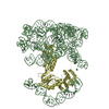 7uimC M: map data used to model this data C: citing same article ( |
|---|---|
| Similar structure data |

-Links
 PDBj
PDBj


- Assembly
| Deposited unit | 
|
|---|---|
| 1 |
|
-Components
-RNA chain / Protein / DNA chain , 3 types, 3 molecules BDA
| #1: RNA chain | Mass: 206779.859 Da / Num. of mol.: 1 / Source method: obtained synthetically / Source: (synth.) [Eubacterium] rectale (bacteria) |
|---|---|
| #2: Protein | Mass: 49083.914 Da / Num. of mol.: 1 Source method: isolated from a genetically manipulated source Source: (gene. exp.) [Eubacterium] rectale (bacteria) / Gene: ltrA_2, ltrA, ERS852417_00966, FYL37_05080 / Production host: References: UniProt: A0A173ZME3, RNA-directed DNA polymerase |
| #3: DNA chain | Mass: 11403.316 Da / Num. of mol.: 1 / Source method: obtained synthetically / Source: (synth.) [Eubacterium] rectale (bacteria) |
-Non-polymers , 3 types, 54 molecules 


| #4: Chemical | ChemComp-MG /  Mass: 24.305 Da / Num. of mol.: 28 / Source method: obtained synthetically / Formula: Mg / Feature type: SUBJECT OF INVESTIGATION Mass: 24.305 Da / Num. of mol.: 28 / Source method: obtained synthetically / Formula: Mg / Feature type: SUBJECT OF INVESTIGATION#5: Chemical |  Mass: 18.038 Da / Num. of mol.: 2 / Source method: obtained synthetically / Formula: H4N / Feature type: SUBJECT OF INVESTIGATION Mass: 18.038 Da / Num. of mol.: 2 / Source method: obtained synthetically / Formula: H4N / Feature type: SUBJECT OF INVESTIGATION#6: Water | ChemComp-HOH / | Mass: 18.015 Da / Num. of mol.: 24 / Source method: isolated from a natural source / Formula: H2O |
|---|
-Details
| Has ligand of interest | Y |
|---|
-Experimental details
-Experiment
| Experiment | Method: ELECTRON MICROSCOPY |
|---|---|
| EM experiment | Aggregation state: PARTICLE / 3D reconstruction method: single particle reconstruction |
- Sample preparation
| Component | Name: Ternary complex of RNA, protein and DNA / Type: COMPLEX / Entity ID: #1-#3 / Source: RECOMBINANT |
|---|---|
| Molecular weight | Value: 0.260 MDa / Experimental value: NO |
| Source (natural) | Organism: [Eubacterium] rectale (bacteria) |
| Source (recombinant) | Organism: |
| Buffer solution | pH: 7.5 |
| Specimen | Embedding applied: NO / Shadowing applied: NO / Staining applied: NO / Vitrification applied: YES |
| Specimen support | Grid material: COPPER / Grid mesh size: 300 divisions/in. / Grid type: Quantifoil R1.2/1.3 |
| Vitrification | Instrument: FEI VITROBOT MARK IV / Cryogen name: ETHANE / Humidity: 100 % / Chamber temperature: 297 K |
- Electron microscopy imaging
| Experimental equipment |  Model: Titan Krios / Image courtesy: FEI Company |
|---|---|
| Microscopy | Model: FEI TITAN KRIOS |
| Electron gun | Electron source:  FIELD EMISSION GUN / Accelerating voltage: 300 kV / Illumination mode: FLOOD BEAM FIELD EMISSION GUN / Accelerating voltage: 300 kV / Illumination mode: FLOOD BEAM |
| Electron lens | Mode: BRIGHT FIELD / Nominal defocus max: 2500 nm / Nominal defocus min: 500 nm |
| Specimen holder | Cryogen: NITROGEN |
| Image recording | Electron dose: 50.5 e/Å2 / Film or detector model: GATAN K3 (6k x 4k) |
- Processing
| EM software |
| ||||||||||||||||||||||||
|---|---|---|---|---|---|---|---|---|---|---|---|---|---|---|---|---|---|---|---|---|---|---|---|---|---|
| CTF correction | Type: PHASE FLIPPING AND AMPLITUDE CORRECTION | ||||||||||||||||||||||||
| 3D reconstruction | Resolution: 2.8 Å / Resolution method: FSC 0.143 CUT-OFF / Num. of particles: 914099 / Symmetry type: POINT |
