 Movie
Movie Controller

[English] 日本語
 Yorodumi
- PDB-7uen: Genetic and structural basis of the human anti-alpha-galactosyl a... -
+ Open data

- Basic information
| Entry | Database: PDB / ID: 7uen | |||||||||
|---|---|---|---|---|---|---|---|---|---|---|
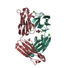
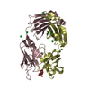
| Title | Genetic and structural basis of the human anti-alpha-galactosyl antibody response | |||||||||
 Components Components | (M86 antibody Fab ...) x 2 | |||||||||
 Keywords Keywords | IMMUNE SYSTEM / alpha-galactosyl / antibody / anti-alpha-gal / immune response / M86 | |||||||||
| Function / homology | Immunoglobulins / Immunoglobulin-like / Sandwich / Mainly Beta / : / PHOSPHATE ION Function and homology information Function and homology information | |||||||||
| Biological species |  | |||||||||
| Method |  X-RAY DIFFRACTION / SYNCHROTRON / MOLECULAR REPLACEMENT / molecular replacement / Resolution: 1.55 Å X-RAY DIFFRACTION / SYNCHROTRON / MOLECULAR REPLACEMENT / molecular replacement / Resolution: 1.55 Å | |||||||||
 Authors Authors | Langley, D.B. / Christ, D. | |||||||||
| Funding support |  Australia, 1items Australia, 1items
| |||||||||
 Citation Citation | Journal: Proc.Natl.Acad.Sci.USA / Year: 2022 Title: Genetic and structural basis of the human anti-alpha-galactosyl antibody response. Authors: Langley, D.B. / Schofield, P. / Nevoltris, D. / Jackson, J. / Jackson, K.J.L. / Peters, T.J. / Burk, M. / Matthews, J.M. / Basten, A. / Goodnow, C.C. / van Nunen, S. / Reed, J.H. / Christ, D. | |||||||||
| History |
|
- Structure visualization
| Structure viewer | Molecule: MolmilJmol/JSmol |
|---|
- Downloads & links
-Download
| PDBx/mmCIF format | 7uen.cif.gz | 353.9 KB | Display | PDBx/mmCIF format |
|---|---|---|---|---|
| PDB format | pdb7uen.ent.gz | 287.4 KB | Display | PDB format |
| PDBx/mmJSON format | 7uen.json.gz | Tree view | PDBx/mmJSON format | |
| Others |  Other downloads Other downloads |
-Validation report
| Arichive directory | https://data.pdbj.org/pub/pdb/validation_reports/ue/7uenftp://data.pdbj.org/pub/pdb/validation_reports/ue/7uen | HTTPS FTP |
|---|
-Related structure data
-Links
 PDBj
PDBj

- Assembly
| Deposited unit | 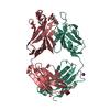
| |||||||||||||||
|---|---|---|---|---|---|---|---|---|---|---|---|---|---|---|---|---|
| 1 |
| |||||||||||||||
| Unit cell |
| |||||||||||||||
| Components on special symmetry positions |
|
-Components
-Antibody , 2 types, 2 molecules LH
| #1: Antibody | Mass: 24065.814 Da / Num. of mol.: 1 Source method: isolated from a genetically manipulated source Source: (gene. exp.)  Homo sapiens (human) Homo sapiens (human) |
|---|---|
| #2: Antibody | Mass: 25106.025 Da / Num. of mol.: 1 Source method: isolated from a genetically manipulated source Source: (gene. exp.) Homo sapiens (human) |
-Sugars , 1 types, 1 molecules
| #3: Polysaccharide | alpha-D-galactopyranose-(1-3)-beta-D-galactopyranose Source method: isolated from a genetically manipulated source |
|---|
-Non-polymers , 3 types, 349 molecules 


| #4: Chemical | ChemComp-K /  Mass: 39.098 Da / Num. of mol.: 1 / Source method: obtained synthetically / Formula: K Mass: 39.098 Da / Num. of mol.: 1 / Source method: obtained synthetically / Formula: K |
|---|---|
| #5: Chemical | ChemComp-PO4 /  Mass: 94.971 Da / Num. of mol.: 1 / Source method: obtained synthetically / Formula: PO4 Mass: 94.971 Da / Num. of mol.: 1 / Source method: obtained synthetically / Formula: PO4 |
| #6: Water | ChemComp-HOH / Mass: 18.015 Da / Num. of mol.: 347 / Source method: isolated from a natural source / Formula: H2O |
-Details
| Has ligand of interest | Y |
|---|---|
| Has protein modification | Y |
-Experimental details
-Experiment
| Experiment | Method: X-RAY DIFFRACTION / Number of used crystals: 1 |
|---|
- Sample preparation
| Crystal | Density Matthews: 2.36 Å3/Da / Density % sol: 47 % |
|---|---|
| Crystal grow | Temperature: 293 K / Method: vapor diffusion, hanging drop / pH: 8 Details: Equal volumes of a protein (in 25 mM Tris (pH 8.0), 100 mM NaCl) and well solution (1.5 M NaH2PO4.H2O.K2HPO4, pH 8.0) were combined. |
-Data collection
| Diffraction | Mean temperature: 100 K / Serial crystal experiment: N | ||||||||||||||||||||||||||||||
|---|---|---|---|---|---|---|---|---|---|---|---|---|---|---|---|---|---|---|---|---|---|---|---|---|---|---|---|---|---|---|---|
| Diffraction source | Source: SYNCHROTRON / Site: Australian Synchrotron / Beamline: MX2 / Wavelength: 0.9537 Å | ||||||||||||||||||||||||||||||
| Detector | Type: DECTRIS EIGER X 16M / Detector: PIXEL / Date: Apr 11, 2017 | ||||||||||||||||||||||||||||||
| Radiation | Protocol: SINGLE WAVELENGTH / Monochromatic (M) / Laue (L): M / Scattering type: x-ray | ||||||||||||||||||||||||||||||
| Radiation wavelength | Wavelength: 0.9537 Å / Relative weight: 1 | ||||||||||||||||||||||||||||||
| Reflection | Resolution: 1.55→38.31 Å / Num. obs: 67630 / % possible obs: 100 % / Redundancy: 18.9 % / CC1/2: 0.999 / Rmerge(I) obs: 0.091 / Rpim(I) all: 0.021 / Rrim(I) all: 0.093 / Net I/σ(I): 17.2 / Num. measured all: 1276211 / Scaling rejects: 224 | ||||||||||||||||||||||||||||||
| Reflection shell | Diffraction-ID: 1
|
-Phasing
| Phasing | Method: molecular replacement | |||||||||
|---|---|---|---|---|---|---|---|---|---|---|
| Phasing MR | Model details: Phaser MODE: MR_AUTO
|
- Processing
| Software |
| |||||||||||||||||||||||||||||||||||||||||||||||||||||||||||||||||
|---|---|---|---|---|---|---|---|---|---|---|---|---|---|---|---|---|---|---|---|---|---|---|---|---|---|---|---|---|---|---|---|---|---|---|---|---|---|---|---|---|---|---|---|---|---|---|---|---|---|---|---|---|---|---|---|---|---|---|---|---|---|---|---|---|---|---|
| Refinement | Method to determine structure: MOLECULAR REPLACEMENT Starting model: generic Fab Resolution: 1.55→38.31 Å / Cor.coef. Fo:Fc: 0.981 / Cor.coef. Fo:Fc free: 0.969 / SU B: 3.111 / SU ML: 0.048 / SU R Cruickshank DPI: 0.0708 / Cross valid method: THROUGHOUT / σ(F): 0 / ESU R: 0.071 / ESU R Free: 0.067 / Stereochemistry target values: MAXIMUM LIKELIHOOD Details: HYDROGENS HAVE BEEN ADDED IN THE RIDING POSITIONS U VALUES : REFINED INDIVIDUALLY
| |||||||||||||||||||||||||||||||||||||||||||||||||||||||||||||||||
| Solvent computation | Ion probe radii: 0.8 Å / Shrinkage radii: 0.8 Å / VDW probe radii: 1.2 Å / Solvent model: MASK | |||||||||||||||||||||||||||||||||||||||||||||||||||||||||||||||||
| Displacement parameters | Biso max: 121.68 Å2 / Biso mean: 27.962 Å2 / Biso min: 14.13 Å2
| |||||||||||||||||||||||||||||||||||||||||||||||||||||||||||||||||
| Refinement step | Cycle: final / Resolution: 1.55→38.31 Å
| |||||||||||||||||||||||||||||||||||||||||||||||||||||||||||||||||
| Refine LS restraints |
| |||||||||||||||||||||||||||||||||||||||||||||||||||||||||||||||||
| LS refinement shell | Resolution: 1.55→1.59 Å / Rfactor Rfree error: 0 / Total num. of bins used: 20
|
