 Movie
Movie Controller

[English] 日本語
 Yorodumi
- PDB-7uem: Genomic and structural basis for the human anti-alpha-galactosyl ... -
+ Open data

- Basic information
| Entry | Database: PDB / ID: 7uem | |||||||||
|---|---|---|---|---|---|---|---|---|---|---|
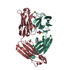
| Title | Genomic and structural basis for the human anti-alpha-galactosyl antibody response | |||||||||
 Components Components |
| |||||||||
 Keywords Keywords | IMMUNE SYSTEM / alpha-galactosyl / antibody / anti-alpha-gal / immune response / M86 | |||||||||
| Function / homology | Immunoglobulins / Immunoglobulin-like / Sandwich / Mainly Beta Function and homology information Function and homology information | |||||||||
| Biological species |  Homo sapiens (human) Homo sapiens (human) | |||||||||
| Method |  X-RAY DIFFRACTION / SYNCHROTRON / MOLECULAR REPLACEMENT / molecular replacement / Resolution: 2.314 Å X-RAY DIFFRACTION / SYNCHROTRON / MOLECULAR REPLACEMENT / molecular replacement / Resolution: 2.314 Å | |||||||||
 Authors Authors | Langley, D.B. / Christ, D. | |||||||||
| Funding support |  Australia, 1items Australia, 1items
| |||||||||
 Citation Citation | Journal: Proc.Natl.Acad.Sci.USA / Year: 2022 Title: Genetic and structural basis of the human anti-alpha-galactosyl antibody response. Authors: Langley, D.B. / Schofield, P. / Nevoltris, D. / Jackson, J. / Jackson, K.J.L. / Peters, T.J. / Burk, M. / Matthews, J.M. / Basten, A. / Goodnow, C.C. / van Nunen, S. / Reed, J.H. / Christ, D. | |||||||||
| History |
|
- Structure visualization
| Structure viewer | Molecule: MolmilJmol/JSmol |
|---|
- Downloads & links
-Download
| PDBx/mmCIF format | 7uem.cif.gz | 346.1 KB | Display | PDBx/mmCIF format |
|---|---|---|---|---|
| PDB format | pdb7uem.ent.gz | 279.9 KB | Display | PDB format |
| PDBx/mmJSON format | 7uem.json.gz | Tree view | PDBx/mmJSON format | |
| Others |  Other downloads Other downloads |
-Validation report
| Arichive directory | https://data.pdbj.org/pub/pdb/validation_reports/ue/7uemftp://data.pdbj.org/pub/pdb/validation_reports/ue/7uem | HTTPS FTP |
|---|
-Related structure data

-Links
 PDBj
PDBj

- Assembly
| Deposited unit | 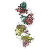
| |||||||||
|---|---|---|---|---|---|---|---|---|---|---|
| 1 | 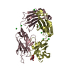
| |||||||||
| 2 | 
| |||||||||
| Unit cell |
| |||||||||
| Components on special symmetry positions |
|
-Components
| #1: Antibody | Mass: 23999.592 Da / Num. of mol.: 2 Source method: isolated from a genetically manipulated source Source: (gene. exp.) Homo sapiens (human) / Cell line (production host): HEK293 / Production host: Homo sapiens (human)#2: Antibody | Mass: 24469.309 Da / Num. of mol.: 2 Source method: isolated from a genetically manipulated source Source: (gene. exp.) Homo sapiens (human) / Cell line (production host): HEK293 / Production host: Homo sapiens (human)#3: Polysaccharide | Source method: isolated from a genetically manipulated source #4: Chemical | ChemComp-CL /   Mass: 35.453 Da / Num. of mol.: 15 / Source method: obtained synthetically / Formula: Cl Mass: 35.453 Da / Num. of mol.: 15 / Source method: obtained synthetically / Formula: Cl#5: Water | ChemComp-HOH / |  Mass: 18.015 Da / Num. of mol.: 320 / Source method: isolated from a natural source / Formula: H2O Mass: 18.015 Da / Num. of mol.: 320 / Source method: isolated from a natural source / Formula: H2OHas ligand of interest | Y | Has protein modification | Y | |
|---|
-Experimental details
-Experiment
| Experiment | Method: X-RAY DIFFRACTION / Number of used crystals: 1 |
|---|
- Sample preparation
| Crystal | Density Matthews: 2.24 Å3/Da / Density % sol: 43 % |
|---|---|
| Crystal grow | Temperature: 293 K / Method: vapor diffusion, sitting drop / pH: 4 Details: Equal volumes of protein (in 25 mM Tris, 100 mM NaCl) were combined with well solution (1 M LiCl2, 100 mM citrate (pH 4.0), 20% (w/v) PEG6000 |
-Data collection
| Diffraction | Mean temperature: 100 K / Serial crystal experiment: N | ||||||||||||||||||||||||||||||
|---|---|---|---|---|---|---|---|---|---|---|---|---|---|---|---|---|---|---|---|---|---|---|---|---|---|---|---|---|---|---|---|
| Diffraction source | Source: SYNCHROTRON / Site: Australian Synchrotron / Beamline: MX2 / Wavelength: 0.9537 Å | ||||||||||||||||||||||||||||||
| Detector | Type: DECTRIS EIGER X 16M / Detector: PIXEL / Date: Aug 24, 2018 | ||||||||||||||||||||||||||||||
| Radiation | Protocol: SINGLE WAVELENGTH / Monochromatic (M) / Laue (L): M / Scattering type: x-ray | ||||||||||||||||||||||||||||||
| Radiation wavelength | Wavelength: 0.9537 Å / Relative weight: 1 | ||||||||||||||||||||||||||||||
| Reflection | Resolution: 2.31→43.46 Å / Num. obs: 37114 / % possible obs: 98.6 % / Redundancy: 5.8 % / Biso Wilson estimate: 33.28 Å2 / CC1/2: 0.995 / Rmerge(I) obs: 0.112 / Rpim(I) all: 0.05 / Rrim(I) all: 0.123 / Net I/σ(I): 10 / Num. measured all: 215453 / Scaling rejects: 24 | ||||||||||||||||||||||||||||||
| Reflection shell | Diffraction-ID: 1
|
-Phasing
| Phasing | Method: molecular replacement | |||||||||
|---|---|---|---|---|---|---|---|---|---|---|
| Phasing MR | Model details: Phaser MODE: MR_AUTO
|
- Processing
| Software |
| ||||||||||||||||||||||||||||||||||||||||||||||||||||||||||||||||||||||||||||||||||||
|---|---|---|---|---|---|---|---|---|---|---|---|---|---|---|---|---|---|---|---|---|---|---|---|---|---|---|---|---|---|---|---|---|---|---|---|---|---|---|---|---|---|---|---|---|---|---|---|---|---|---|---|---|---|---|---|---|---|---|---|---|---|---|---|---|---|---|---|---|---|---|---|---|---|---|---|---|---|---|---|---|---|---|---|---|---|
| Refinement | Method to determine structure: MOLECULAR REPLACEMENT Starting model: generic Fab Resolution: 2.314→42.974 Å / SU ML: 0.3 / Cross valid method: THROUGHOUT / σ(F): 1.33 / Phase error: 26.07 / Stereochemistry target values: ML
| ||||||||||||||||||||||||||||||||||||||||||||||||||||||||||||||||||||||||||||||||||||
| Solvent computation | Shrinkage radii: 0.9 Å / VDW probe radii: 1.11 Å / Solvent model: FLAT BULK SOLVENT MODEL | ||||||||||||||||||||||||||||||||||||||||||||||||||||||||||||||||||||||||||||||||||||
| Displacement parameters | Biso max: 76.32 Å2 / Biso mean: 37.0057 Å2 / Biso min: 20.13 Å2 | ||||||||||||||||||||||||||||||||||||||||||||||||||||||||||||||||||||||||||||||||||||
| Refinement step | Cycle: final / Resolution: 2.314→42.974 Å
| ||||||||||||||||||||||||||||||||||||||||||||||||||||||||||||||||||||||||||||||||||||
| Refine LS restraints |
| ||||||||||||||||||||||||||||||||||||||||||||||||||||||||||||||||||||||||||||||||||||
| LS refinement shell | Refine-ID: X-RAY DIFFRACTION / Rfactor Rfree error: 0
| ||||||||||||||||||||||||||||||||||||||||||||||||||||||||||||||||||||||||||||||||||||
| Refinement TLS params. | Method: refined / Origin x: 21.264 Å / Origin y: 16.8824 Å / Origin z: 38.0035 Å
| ||||||||||||||||||||||||||||||||||||||||||||||||||||||||||||||||||||||||||||||||||||
| Refinement TLS group |
|
