| 登録情報 | データベース: PDB / ID: 7udj
|
|---|
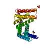
| タイトル | Crystal structure of designed helical repeat protein RPB_PEW3_R4 bound to PAWx4 peptide |
|---|
 要素 要素 | - 4xPAW peptide
- De novo designed helical repeat protein RPB_PEW3_R4
|
|---|
 キーワード キーワード | DE NOVO PROTEIN / Synthetic protein / designed helical repeat protein / peptide binder |
|---|
| 生物種 | synthetic construct (人工物) |
|---|
| 手法 |  X線回折 / シンクロトロン / 分子置換 / 解像度: 2.7 Å X線回折 / シンクロトロン / 分子置換 / 解像度: 2.7 Å |
|---|
 データ登録者 データ登録者 | Redler, R.L. / Chang, Y. / Bhabha, G. / Ekiert, D. |
|---|
| 資金援助 |  米国, 1件 米国, 1件 | 組織 | 認可番号 | 国 |
|---|
| National Institutes of Health/National Institute of General Medical Sciences (NIH/NIGMS) | GM128777 | 米国 |
|
|---|
 引用 引用 | ジャーナル: Nature / 年: 2023
タイトル: De novo design of modular peptide-binding proteins by superhelical matching.
著者: Wu, K. / Bai, H. / Chang, Y.T. / Redler, R. / McNally, K.E. / Sheffler, W. / Brunette, T.J. / Hicks, D.R. / Morgan, T.E. / Stevens, T.J. / Broerman, A. / Goreshnik, I. / DeWitt, M. / Chow, C. ...著者: Wu, K. / Bai, H. / Chang, Y.T. / Redler, R. / McNally, K.E. / Sheffler, W. / Brunette, T.J. / Hicks, D.R. / Morgan, T.E. / Stevens, T.J. / Broerman, A. / Goreshnik, I. / DeWitt, M. / Chow, C.M. / Shen, Y. / Stewart, L. / Derivery, E. / Silva, D.A. / Bhabha, G. / Ekiert, D.C. / Baker, D. |
|---|
| 履歴 | | 登録 | 2022年3月20日 | 登録サイト: RCSB / 処理サイト: RCSB |
|---|
| 改定 1.0 | 2023年3月22日 | Provider: repository / タイプ: Initial release |
|---|
| 改定 1.1 | 2023年4月19日 | Group: Database references / カテゴリ: citation / citation_author
Item: _citation.country / _citation.journal_abbrev ..._citation.country / _citation.journal_abbrev / _citation.journal_id_ASTM / _citation.journal_id_CSD / _citation.journal_id_ISSN / _citation.pdbx_database_id_DOI / _citation.pdbx_database_id_PubMed / _citation.title / _citation.year |
|---|
| 改定 1.2 | 2023年5月3日 | Group: Database references / カテゴリ: citation / citation_author
Item: _citation.journal_volume / _citation.page_first ..._citation.journal_volume / _citation.page_first / _citation.page_last / _citation_author.identifier_ORCID |
|---|
| 改定 1.3 | 2024年4月3日 | Group: Data collection / Refinement description
カテゴリ: chem_comp_atom / chem_comp_bond / pdbx_initial_refinement_model |
|---|
|
|---|
 ムービー
ムービー コントローラー


 データを開く
 基本情報
基本情報 構造の表示
構造の表示 Molmil
 ダウンロードとリンク
 その他のダウンロード


 PDBj
PDBj
 集合体
集合体

 試料調製
試料調製 解析
解析