 Movie
Movie Controller


+ Open data

- Basic information
| Entry | Database: PDB / ID: 7uda | ||||||
|---|---|---|---|---|---|---|---|
| Title | Structure of the EstG | ||||||
 Components Components | Beta-lactamase domain-containing protein | ||||||
 Keywords Keywords | HYDROLASE / alpha/beta fold | ||||||
| Function / homology | : / Beta-lactamase-related / Beta-lactamase / Beta-lactamase/transpeptidase-like / Beta-lactamase-related domain-containing protein Function and homology information Function and homology information | ||||||
| Biological species |  Caulobacter vibrioides (bacteria) Caulobacter vibrioides (bacteria) | ||||||
| Method |  X-RAY DIFFRACTION / SYNCHROTRON / MOLECULAR REPLACEMENT / Resolution: 2.47 Å X-RAY DIFFRACTION / SYNCHROTRON / MOLECULAR REPLACEMENT / Resolution: 2.47 Å | ||||||
 Authors Authors | Chen, Z. / Gabelli, S.B. | ||||||
| Funding support |  United States, 1items United States, 1items
| ||||||
 Citation Citation | Journal: Curr.Biol. / Year: 2023 Title: EstG is a novel esterase required for cell envelope integrity in Caulobacter. Authors: Daitch, A.K. / Orsburn, B.C. / Chen, Z. / Alvarez, L. / Eberhard, C.D. / Sundararajan, K. / Zeinert, R. / Kreitler, D.F. / Jakoncic, J. / Chien, P. / Cava, F. / Gabelli, S.B. / Goley, E.D. | ||||||
| History |
|
- Structure visualization
| Structure viewer | Molecule: MolmilJmol/JSmol |
|---|
- Downloads & links
-Download
| PDBx/mmCIF format | 7uda.cif.gz | 93.2 KB | Display | PDBx/mmCIF format |
|---|---|---|---|---|
| PDB format | pdb7uda.ent.gz | 68.2 KB | Display | PDB format |
| PDBx/mmJSON format | 7uda.json.gz | Tree view | PDBx/mmJSON format | |
| Others |  Other downloads Other downloads |
-Validation report
| Arichive directory | https://data.pdbj.org/pub/pdb/validation_reports/ud/7udaftp://data.pdbj.org/pub/pdb/validation_reports/ud/7uda | HTTPS FTP |
|---|
-Related structure data
| Related structure data | 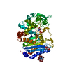 7u1bC  7u1cC  1ci8S C: citing same article ( S: Starting model for refinement |
|---|---|
| Similar structure data |
-Links
 PDBj
PDBj
- Assembly
| Deposited unit | 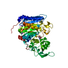
| ||||||||
|---|---|---|---|---|---|---|---|---|---|
| 1 |
| ||||||||
| Unit cell |
|
-Components
| #1: Protein | Mass: 49224.500 Da / Num. of mol.: 1 Fragment: estG, Esterase for Stress Tolerance acting on glucans Source method: isolated from a genetically manipulated source Source: (gene. exp.) Caulobacter vibrioides (bacteria) / Plasmid: pEG1622 / Production host: | ||||
|---|---|---|---|---|---|
| #2: Chemical | ChemComp-TRS /   Mass: 122.143 Da / Num. of mol.: 1 / Source method: obtained synthetically / Formula: C4H12NO3 / Comment: pH buffer*YM Mass: 122.143 Da / Num. of mol.: 1 / Source method: obtained synthetically / Formula: C4H12NO3 / Comment: pH buffer*YM | ||||
| #3: Chemical | ChemComp-NA /   Mass: 22.990 Da / Num. of mol.: 4 / Source method: obtained synthetically / Formula: Na Mass: 22.990 Da / Num. of mol.: 4 / Source method: obtained synthetically / Formula: Na#4: Water | ChemComp-HOH / |  Mass: 18.015 Da / Num. of mol.: 31 / Source method: isolated from a natural source / Formula: H2O Mass: 18.015 Da / Num. of mol.: 31 / Source method: isolated from a natural source / Formula: H2OHas ligand of interest | N | |
-Experimental details
-Experiment
| Experiment | Method: X-RAY DIFFRACTION / Number of used crystals: 1 |
|---|
- Sample preparation
| Crystal | Density Matthews: 3.46 Å3/Da / Density % sol: 64.41 % / Mosaicity: 0.13 ° |
|---|---|
| Crystal grow | Temperature: 298 K / Method: vapor diffusion / pH: 8.5 Details: 20% PEG500 MME, 10% PEG20000, 0.1 M Tris/Bicine pH 8.5 and 90 mM mixture of sodium nitrate, sodium phosphate dibasic and ammonium sulfate |
-Data collection
| Diffraction | Mean temperature: 277 K / Serial crystal experiment: N | |||||||||||||||||||||||||||
|---|---|---|---|---|---|---|---|---|---|---|---|---|---|---|---|---|---|---|---|---|---|---|---|---|---|---|---|---|
| Diffraction source | Source: SYNCHROTRON / Type: OTHER / Wavelength: 0.9793 Å | |||||||||||||||||||||||||||
| Detector | Type: DECTRIS EIGER X 16M / Detector: PIXEL / Date: May 27, 2021 | |||||||||||||||||||||||||||
| Radiation | Protocol: SINGLE WAVELENGTH / Monochromatic (M) / Laue (L): M / Scattering type: x-ray | |||||||||||||||||||||||||||
| Radiation wavelength | Wavelength: 0.9793 Å / Relative weight: 1 | |||||||||||||||||||||||||||
| Reflection | Resolution: 2.47→29.62 Å / Num. obs: 24085 / % possible obs: 99 % / Redundancy: 6.7 % / CC1/2: 0.996 / Rmerge(I) obs: 0.108 / Rpim(I) all: 0.045 / Rrim(I) all: 0.117 / Net I/σ(I): 10.9 | |||||||||||||||||||||||||||
| Reflection shell | Diffraction-ID: 1 / Redundancy: 6.5 %
|
- Processing
| Software |
| ||||||||||||||||||||||||||||||||||||||||||||||||||||||||||||
|---|---|---|---|---|---|---|---|---|---|---|---|---|---|---|---|---|---|---|---|---|---|---|---|---|---|---|---|---|---|---|---|---|---|---|---|---|---|---|---|---|---|---|---|---|---|---|---|---|---|---|---|---|---|---|---|---|---|---|---|---|---|
| Refinement | Method to determine structure: MOLECULAR REPLACEMENT Starting model: 1ci8 Resolution: 2.47→29.62 Å / Cor.coef. Fo:Fc: 0.96 / Cor.coef. Fo:Fc free: 0.939 / WRfactor Rfree: 0.2159 / WRfactor Rwork: 0.1752 / FOM work R set: 0.8406 / SU B: 6.945 / SU ML: 0.154 / SU R Cruickshank DPI: 0.2452 / SU Rfree: 0.203 / Cross valid method: THROUGHOUT / σ(F): 0 / ESU R: 0.245 / ESU R Free: 0.203 / Stereochemistry target values: MAXIMUM LIKELIHOOD Details: HYDROGENS HAVE BEEN ADDED IN THE RIDING POSITIONS U VALUES : REFINED INDIVIDUALLY
| ||||||||||||||||||||||||||||||||||||||||||||||||||||||||||||
| Solvent computation | Ion probe radii: 0.8 Å / Shrinkage radii: 0.8 Å / VDW probe radii: 1.2 Å / Solvent model: MASK | ||||||||||||||||||||||||||||||||||||||||||||||||||||||||||||
| Displacement parameters | Biso max: 187.25 Å2 / Biso mean: 52.356 Å2 / Biso min: 23.82 Å2
| ||||||||||||||||||||||||||||||||||||||||||||||||||||||||||||
| Refinement step | Cycle: final / Resolution: 2.47→29.62 Å
| ||||||||||||||||||||||||||||||||||||||||||||||||||||||||||||
| Refine LS restraints |
| ||||||||||||||||||||||||||||||||||||||||||||||||||||||||||||
| LS refinement shell | Resolution: 2.474→2.538 Å / Rfactor Rfree error: 0 / Total num. of bins used: 20
|
