 Movie
Movie Controller

[English] 日本語
 Yorodumi
- PDB-7u9c: Crystal Structure of the wild type Escherichia coli Pyridoxal 5'-... -
+ Open data

- Basic information
| Entry | Database: PDB / ID: 7u9c | |||||||||
|---|---|---|---|---|---|---|---|---|---|---|
| Title | Crystal Structure of the wild type Escherichia coli Pyridoxal 5'-phosphate homeostasis protein (YGGS) | |||||||||
 Components Components | Pyridoxal phosphate homeostasis protein | |||||||||
 Keywords Keywords | PROTEIN TRANSPORT / PLP-binding protein / Vitamin B6 / PLP homeostasis | |||||||||
| Function / homology | Uncharacterized protein family UPF0001 signature. / Pyridoxal phosphate homeostasis protein / Alanine racemase, N-terminal / Alanine racemase, N-terminal domain / PLP-binding barrel / pyridoxal phosphate binding / PYRIDOXAL-5'-PHOSPHATE / PHOSPHATE ION / Pyridoxal phosphate homeostasis protein Function and homology information Function and homology information | |||||||||
| Biological species |  | |||||||||
| Method |  X-RAY DIFFRACTION / MOLECULAR REPLACEMENT / molecular replacement / Resolution: 2.1 Å X-RAY DIFFRACTION / MOLECULAR REPLACEMENT / molecular replacement / Resolution: 2.1 Å | |||||||||
 Authors Authors | Donkor, A.K. / Ghatge, M.S. / Safo, M.K. / Musayev, F.N. | |||||||||
| Funding support |  United States, 2items United States, 2items
| |||||||||
 Citation Citation | Journal: Protein Sci. / Year: 2022 Title: Characterization of the Escherichia coli pyridoxal 5'-phosphate homeostasis protein (YggS): Role of lysine residues in PLP binding and protein stability. Authors: Tramonti, A. / Ghatge, M.S. / Babor, J.T. / Musayev, F.N. / di Salvo, M.L. / Barile, A. / Colotti, G. / Giorgi, A. / Paredes, S.D. / Donkor, A.K. / Al Mughram, M.H. / de Crecy-Lagard, V. / ...Authors: Tramonti, A. / Ghatge, M.S. / Babor, J.T. / Musayev, F.N. / di Salvo, M.L. / Barile, A. / Colotti, G. / Giorgi, A. / Paredes, S.D. / Donkor, A.K. / Al Mughram, M.H. / de Crecy-Lagard, V. / Safo, M.K. / Contestabile, R. | |||||||||
| History |
|
- Structure visualization
| Structure viewer | Molecule: MolmilJmol/JSmol |
|---|
- Downloads & links
-Download
| PDBx/mmCIF format | 7u9c.cif.gz | 64.9 KB | Display | PDBx/mmCIF format |
|---|---|---|---|---|
| PDB format | pdb7u9c.ent.gz | 45.2 KB | Display | PDB format |
| PDBx/mmJSON format | 7u9c.json.gz | Tree view | PDBx/mmJSON format | |
| Others |  Other downloads Other downloads |
-Validation report
| Arichive directory | https://data.pdbj.org/pub/pdb/validation_reports/u9/7u9cftp://data.pdbj.org/pub/pdb/validation_reports/u9/7u9c | HTTPS FTP |
|---|
-Related structure data
| Related structure data | 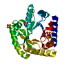 7u9hC  7uatC  7uauC 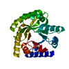 7uaxC  7ub4C  7ub8C 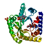 7ubpC 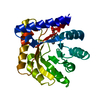 7ubqC  1w8gS S: Starting model for refinement C: citing same article ( |
|---|---|
| Similar structure data |
-Links
 PDBj
PDBj- Assembly
| Deposited unit | 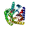
| |||||||||
|---|---|---|---|---|---|---|---|---|---|---|
| 1 |
| |||||||||
| Unit cell |
| |||||||||
| Components on special symmetry positions |
|
-Components
| #1: Protein | Mass: 25820.441 Da / Num. of mol.: 1 Source method: isolated from a genetically manipulated source Source: (gene. exp.) | ||||||
|---|---|---|---|---|---|---|---|
| #2: Chemical |   Mass: 247.142 Da / Num. of mol.: 2 / Source method: obtained synthetically / Formula: C8H10NO6P / Feature type: SUBJECT OF INVESTIGATION Mass: 247.142 Da / Num. of mol.: 2 / Source method: obtained synthetically / Formula: C8H10NO6P / Feature type: SUBJECT OF INVESTIGATION#3: Chemical | ChemComp-PO4 / |   Mass: 94.971 Da / Num. of mol.: 1 / Source method: obtained synthetically / Formula: PO4 / Feature type: SUBJECT OF INVESTIGATION Mass: 94.971 Da / Num. of mol.: 1 / Source method: obtained synthetically / Formula: PO4 / Feature type: SUBJECT OF INVESTIGATION#4: Water | ChemComp-HOH / |  Mass: 18.015 Da / Num. of mol.: 188 / Source method: isolated from a natural source / Formula: H2O Mass: 18.015 Da / Num. of mol.: 188 / Source method: isolated from a natural source / Formula: H2OHas ligand of interest | Y | |
-Experimental details
-Experiment
| Experiment | Method: X-RAY DIFFRACTION / Number of used crystals: 1 |
|---|
- Sample preparation
| Crystal | Density Matthews: 3.1 Å3/Da / Density % sol: 60.36 % |
|---|---|
| Crystal grow | Temperature: 293 K / Method: vapor diffusion, hanging drop / pH: 7.5 / Details: 0.1M HEPES/NaOH pH 7.5, 0.8 NaH2PO4/0.8M KH2PO4 |
-Data collection
| Diffraction | Mean temperature: 100 K / Serial crystal experiment: N | ||||||||||||||||||||||||
|---|---|---|---|---|---|---|---|---|---|---|---|---|---|---|---|---|---|---|---|---|---|---|---|---|---|
| Diffraction source | Source: ROTATING ANODE / Type: RIGAKU MICROMAX-007 HF / Wavelength: 1.5406 Å | ||||||||||||||||||||||||
| Detector | Type: DECTRIS EIGER R 4M / Detector: PIXEL / Date: Mar 11, 2021 / Details: VariMax TM-VHF Arc)Sec Confocal Optical System | ||||||||||||||||||||||||
| Radiation | Protocol: SINGLE WAVELENGTH / Monochromatic (M) / Laue (L): M / Scattering type: x-ray | ||||||||||||||||||||||||
| Radiation wavelength | Wavelength: 1.5406 Å / Relative weight: 1 | ||||||||||||||||||||||||
| Reflection | Resolution: 2.1→29.271 Å / Num. obs: 19252 / % possible obs: 100 % / Redundancy: 13.8 % / Biso Wilson estimate: 31.82 Å2 / CC1/2: 1 / Rmerge(I) obs: 0.069 / Rpim(I) all: 0.019 / Rrim(I) all: 0.071 / Net I/σ(I): 30.4 | ||||||||||||||||||||||||
| Reflection shell | Diffraction-ID: 1
|
-Phasing
| Phasing | Method: molecular replacement |
|---|
- Processing
| Software |
| ||||||||||||||||||||||||||||||||||||||||
|---|---|---|---|---|---|---|---|---|---|---|---|---|---|---|---|---|---|---|---|---|---|---|---|---|---|---|---|---|---|---|---|---|---|---|---|---|---|---|---|---|---|
| Refinement | Method to determine structure: MOLECULAR REPLACEMENT Starting model: 1w8g Resolution: 2.1→29.271 Å / SU ML: 0.2 / Cross valid method: THROUGHOUT / σ(F): 1.34 / Phase error: 21.17 / Stereochemistry target values: ML
| ||||||||||||||||||||||||||||||||||||||||
| Solvent computation | Shrinkage radii: 0.9 Å / VDW probe radii: 1.11 Å / Solvent model: FLAT BULK SOLVENT MODEL | ||||||||||||||||||||||||||||||||||||||||
| Displacement parameters | Biso max: 102.49 Å2 / Biso mean: 34.9622 Å2 / Biso min: 17.92 Å2 | ||||||||||||||||||||||||||||||||||||||||
| Refinement step | Cycle: final / Resolution: 2.1→29.271 Å
| ||||||||||||||||||||||||||||||||||||||||
| LS refinement shell | Refine-ID: X-RAY DIFFRACTION / Rfactor Rfree error: 0 / % reflection obs: 100 %
|
