 Movie
Movie Controller


+ Open data

- Basic information
| Entry | Database: PDB / ID: 7u6i | ||||||
|---|---|---|---|---|---|---|---|
| Title | HalB with glycine and succinate | ||||||
 Components Components | Halogenase B | ||||||
 Keywords Keywords | BIOSYNTHETIC PROTEIN | ||||||
| Function / homology | : / Halogenase D / q2cbj1_9rhob like domain / Jelly Rolls / Sandwich / Mainly Beta / GLYCINE / SUCCINIC ACID / ArpA protein Function and homology information Function and homology information | ||||||
| Biological species |  Streptomyces wuyuanensis (bacteria) Streptomyces wuyuanensis (bacteria) | ||||||
| Method |  X-RAY DIFFRACTION / SYNCHROTRON / MOLECULAR REPLACEMENT / Resolution: 2.05 Å X-RAY DIFFRACTION / SYNCHROTRON / MOLECULAR REPLACEMENT / Resolution: 2.05 Å | ||||||
 Authors Authors | Neugebauer, M.E. / Kissman, E.N. / Chang, M.C.Y. | ||||||
| Funding support |  United States, 1items United States, 1items
| ||||||
 Citation Citation | Journal: Proc.Natl.Acad.Sci.USA / Year: 2023 Title: Biocatalytic control of site-selectivity and chain length-selectivity in radical amino acid halogenases. Authors: Kissman, E.N. / Neugebauer, M.E. / Sumida, K.H. / Swenson, C.V. / Sambold, N.A. / Marchand, J.A. / Millar, D.C. / Chang, M.C.Y. | ||||||
| History |
|
- Structure visualization
| Structure viewer | Molecule: MolmilJmol/JSmol |
|---|
- Downloads & links
-Download
| PDBx/mmCIF format | 7u6i.cif.gz | 120.7 KB | Display | PDBx/mmCIF format |
|---|---|---|---|---|
| PDB format | pdb7u6i.ent.gz | 92.6 KB | Display | PDB format |
| PDBx/mmJSON format | 7u6i.json.gz | Tree view | PDBx/mmJSON format | |
| Others |  Other downloads Other downloads |
-Validation report
| Arichive directory | https://data.pdbj.org/pub/pdb/validation_reports/u6/7u6iftp://data.pdbj.org/pub/pdb/validation_reports/u6/7u6i | HTTPS FTP |
|---|
-Related structure data
| Related structure data |  7u6hC 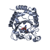 7u6jC  6nieS S: Starting model for refinement C: citing same article ( |
|---|---|
| Similar structure data |
-Links
 PDBj
PDBj

- Assembly
| Deposited unit | 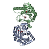
| ||||||||
|---|---|---|---|---|---|---|---|---|---|
| 1 | 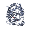
| ||||||||
| 2 | 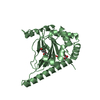
| ||||||||
| Unit cell |
|
-Components
| #1: Protein | Mass: 28632.223 Da / Num. of mol.: 2 Source method: isolated from a genetically manipulated source Source: (gene. exp.) Streptomyces wuyuanensis (bacteria) / Gene: SAMN05444921_127108 / Production host: #2: Chemical |   Mass: 118.088 Da / Num. of mol.: 2 / Source method: obtained synthetically / Formula: C4H6O4 / Feature type: SUBJECT OF INVESTIGATION Mass: 118.088 Da / Num. of mol.: 2 / Source method: obtained synthetically / Formula: C4H6O4 / Feature type: SUBJECT OF INVESTIGATION#3: Chemical | ChemComp-GLY / |   Type: peptide linking / Mass: 75.067 Da / Num. of mol.: 1 / Source method: obtained synthetically / Formula: C2H5NO2 / Feature type: SUBJECT OF INVESTIGATION Type: peptide linking / Mass: 75.067 Da / Num. of mol.: 1 / Source method: obtained synthetically / Formula: C2H5NO2 / Feature type: SUBJECT OF INVESTIGATION#4: Chemical | ChemComp-GOL / |   Mass: 92.094 Da / Num. of mol.: 1 / Source method: obtained synthetically / Formula: C3H8O3 Mass: 92.094 Da / Num. of mol.: 1 / Source method: obtained synthetically / Formula: C3H8O3#5: Water | ChemComp-HOH / |  Mass: 18.015 Da / Num. of mol.: 439 / Source method: isolated from a natural source / Formula: H2O Mass: 18.015 Da / Num. of mol.: 439 / Source method: isolated from a natural source / Formula: H2OHas ligand of interest | Y | |
|---|
-Experimental details
-Experiment
| Experiment | Method: X-RAY DIFFRACTION / Number of used crystals: 1 |
|---|
- Sample preparation
| Crystal | Density Matthews: 3.64 Å3/Da / Density % sol: 66.16 % |
|---|---|
| Crystal grow | Temperature: 291 K / Method: vapor diffusion, hanging drop / pH: 5.2 Details: Equal volumes of protein solution (SwHalB (2.5 mg/ml), lysine (3 mM), alpha-ketoglutarate (3 mM, pH 7)) and reservoir solution (succinate phosphate glycine (SPG) buffer (100 mM, pH 5.2), 18% ...Details: Equal volumes of protein solution (SwHalB (2.5 mg/ml), lysine (3 mM), alpha-ketoglutarate (3 mM, pH 7)) and reservoir solution (succinate phosphate glycine (SPG) buffer (100 mM, pH 5.2), 18% (w/v) PEG 1500) were mixed |
-Data collection
| Diffraction | Mean temperature: 194 K / Serial crystal experiment: N |
|---|---|
| Diffraction source | Source: SYNCHROTRON / Site: ALS / Beamline: 8.3.1 / Wavelength: 1.11 Å |
| Detector | Type: DECTRIS PILATUS3 S 6M / Detector: PIXEL / Date: Mar 28, 2018 |
| Radiation | Protocol: SINGLE WAVELENGTH / Monochromatic (M) / Laue (L): M / Scattering type: x-ray |
| Radiation wavelength | Wavelength: 1.11 Å / Relative weight: 1 |
| Reflection | Resolution: 2.05→143.41 Å / Num. obs: 53826 / % possible obs: 99.8 % / Redundancy: 39.65 % / CC1/2: 1 / Net I/σ(I): 23.9 |
| Reflection shell | Resolution: 2.05→2.1 Å / Redundancy: 6 % / Mean I/σ(I) obs: 1.5 / Num. unique obs: 3989 / CC1/2: 0.706 / % possible all: 97.3 |
- Processing
| Software |
| |||||||||||||||||||||||||||||||||||||||||||||||||||||||||||||||||||||||||||||||||||||||||||||||||||||||||||||||||||||||||||||||||||||||||||||||||||
|---|---|---|---|---|---|---|---|---|---|---|---|---|---|---|---|---|---|---|---|---|---|---|---|---|---|---|---|---|---|---|---|---|---|---|---|---|---|---|---|---|---|---|---|---|---|---|---|---|---|---|---|---|---|---|---|---|---|---|---|---|---|---|---|---|---|---|---|---|---|---|---|---|---|---|---|---|---|---|---|---|---|---|---|---|---|---|---|---|---|---|---|---|---|---|---|---|---|---|---|---|---|---|---|---|---|---|---|---|---|---|---|---|---|---|---|---|---|---|---|---|---|---|---|---|---|---|---|---|---|---|---|---|---|---|---|---|---|---|---|---|---|---|---|---|---|---|---|---|
| Refinement | Method to determine structure: MOLECULAR REPLACEMENT Starting model: 6NIE Resolution: 2.05→82.74 Å / SU ML: 0.27 / Cross valid method: THROUGHOUT / σ(F): 1.33 / Phase error: 26.12 / Stereochemistry target values: ML
| |||||||||||||||||||||||||||||||||||||||||||||||||||||||||||||||||||||||||||||||||||||||||||||||||||||||||||||||||||||||||||||||||||||||||||||||||||
| Solvent computation | Shrinkage radii: 0.9 Å / VDW probe radii: 1.11 Å / Solvent model: FLAT BULK SOLVENT MODEL | |||||||||||||||||||||||||||||||||||||||||||||||||||||||||||||||||||||||||||||||||||||||||||||||||||||||||||||||||||||||||||||||||||||||||||||||||||
| Displacement parameters | Biso max: 112.59 Å2 / Biso mean: 47.3102 Å2 / Biso min: 22.59 Å2 | |||||||||||||||||||||||||||||||||||||||||||||||||||||||||||||||||||||||||||||||||||||||||||||||||||||||||||||||||||||||||||||||||||||||||||||||||||
| Refinement step | Cycle: final / Resolution: 2.05→82.74 Å
| |||||||||||||||||||||||||||||||||||||||||||||||||||||||||||||||||||||||||||||||||||||||||||||||||||||||||||||||||||||||||||||||||||||||||||||||||||
| LS refinement shell | Refine-ID: X-RAY DIFFRACTION / Rfactor Rfree error: 0 / Total num. of bins used: 20
|
