 Movie
Movie Controller


+ Open data

- Basic information
| Entry | Database: PDB / ID: 7u4i | ||||||||||||||||||||||||
|---|---|---|---|---|---|---|---|---|---|---|---|---|---|---|---|---|---|---|---|---|---|---|---|---|---|
| Title | Crystal structure of human GPX4-U46C-R152H in complex with CDS9 | ||||||||||||||||||||||||
 Components Components | Phospholipid hydroperoxide glutathione peroxidase | ||||||||||||||||||||||||
 Keywords Keywords | OXIDOREDUCTASE / GPX4 | ||||||||||||||||||||||||
| Function / homology |  Function and homology information Function and homology informationphospholipid-hydroperoxide glutathione peroxidase / phospholipid-hydroperoxide glutathione peroxidase activity / Synthesis of 12-eicosatetraenoic acid derivatives / Synthesis of 15-eicosatetraenoic acid derivatives / selenium binding / Synthesis of 5-eicosatetraenoic acids / glutathione peroxidase / lipoxygenase pathway / Biosynthesis of aspirin-triggered D-series resolvins / Biosynthesis of E-series 18(R)-resolvins ...phospholipid-hydroperoxide glutathione peroxidase / phospholipid-hydroperoxide glutathione peroxidase activity / Synthesis of 12-eicosatetraenoic acid derivatives / Synthesis of 15-eicosatetraenoic acid derivatives / selenium binding / Synthesis of 5-eicosatetraenoic acids / glutathione peroxidase / lipoxygenase pathway / Biosynthesis of aspirin-triggered D-series resolvins / Biosynthesis of E-series 18(R)-resolvins / Biosynthesis of D-series resolvins / Biosynthesis of E-series 18(S)-resolvins / arachidonate metabolic process / glutathione peroxidase activity / long-chain fatty acid biosynthetic process / dendrite development / negative regulation of ferroptosis / protein polymerization / phospholipid metabolic process / cerebellum development / multicellular organism growth / response to estradiol / nuclear envelope / chromatin organization / response to oxidative stress / spermatogenesis / response to lipopolysaccharide / apoptotic process / protein-containing complex / mitochondrion / extracellular exosome / identical protein binding / nucleus / cytosol Similarity search - Function | ||||||||||||||||||||||||
| Biological species |  Homo sapiens (human) Homo sapiens (human) | ||||||||||||||||||||||||
| Method |  X-RAY DIFFRACTION / SYNCHROTRON / MOLECULAR REPLACEMENT / Resolution: 1.97 Å X-RAY DIFFRACTION / SYNCHROTRON / MOLECULAR REPLACEMENT / Resolution: 1.97 Å | ||||||||||||||||||||||||
 Authors Authors | Forouhar, F. / Liu, H. / Lin, A.J. / Wang, Q. / Polychronidou, V. / Soni, R.K. / Xia, X. / Stockwell, B.R. | ||||||||||||||||||||||||
| Funding support |  United States, 7items United States, 7items
| ||||||||||||||||||||||||
 Citation Citation | Journal: Cell Chem Biol / Year: 2022 Title: Small-molecule allosteric inhibitors of GPX4. Authors: Liu, H. / Forouhar, F. / Lin, A.J. / Wang, Q. / Polychronidou, V. / Soni, R.K. / Xia, X. / Stockwell, B.R. | ||||||||||||||||||||||||
| History |
|
- Structure visualization
| Structure viewer | Molecule: MolmilJmol/JSmol |
|---|
- Downloads & links
-Download
| PDBx/mmCIF format | 7u4i.cif.gz | 152.9 KB | Display | PDBx/mmCIF format |
|---|---|---|---|---|
| PDB format | pdb7u4i.ent.gz | 118.7 KB | Display | PDB format |
| PDBx/mmJSON format | 7u4i.json.gz | Tree view | PDBx/mmJSON format | |
| Others |  Other downloads Other downloads |
-Validation report
| Arichive directory | https://data.pdbj.org/pub/pdb/validation_reports/u4/7u4iftp://data.pdbj.org/pub/pdb/validation_reports/u4/7u4i | HTTPS FTP |
|---|
-Related structure data
| Related structure data | 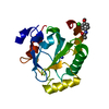 7u4jC  7u4kC 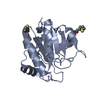 7u4lC  7u4mC  7u4nC  7l8lS S: Starting model for refinement C: citing same article ( |
|---|---|
| Similar structure data |
-Links
 PDBj
PDBj

- Assembly
| Deposited unit | 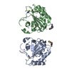
| ||||||||
|---|---|---|---|---|---|---|---|---|---|
| 1 | 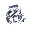
| ||||||||
| 2 | 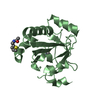
| ||||||||
| Unit cell |
|
-Components
| #1: Protein | Mass: 21869.051 Da / Num. of mol.: 2 / Mutation: U46C, R152H Source method: isolated from a genetically manipulated source Source: (gene. exp.) Homo sapiens (human) / Gene: GPX4 / Production host:  References: UniProt: P36969, phospholipid-hydroperoxide glutathione peroxidase #2: Chemical |   Mass: 234.114 Da / Num. of mol.: 2 / Source method: obtained synthetically / Formula: C7H8BrNOS Mass: 234.114 Da / Num. of mol.: 2 / Source method: obtained synthetically / Formula: C7H8BrNOS#3: Chemical |   Mass: 58.082 Da / Num. of mol.: 2 / Source method: obtained synthetically / Formula: CNS / Feature type: SUBJECT OF INVESTIGATION Mass: 58.082 Da / Num. of mol.: 2 / Source method: obtained synthetically / Formula: CNS / Feature type: SUBJECT OF INVESTIGATION#4: Water | ChemComp-HOH / |  Mass: 18.015 Da / Num. of mol.: 179 / Source method: isolated from a natural source / Formula: H2O Mass: 18.015 Da / Num. of mol.: 179 / Source method: isolated from a natural source / Formula: H2OHas ligand of interest | Y | Has protein modification | Y | |
|---|
-Experimental details
-Experiment
| Experiment | Method: X-RAY DIFFRACTION / Number of used crystals: 1 |
|---|
- Sample preparation
| Crystal | Density Matthews: 2.18 Å3/Da / Density % sol: 43.47 % |
|---|---|
| Crystal grow | Temperature: 291 K / Method: microbatch / pH: 7 / Details: 0.2 M potassium thiocyanate and 20% (w/v) PEG 3350 |
-Data collection
| Diffraction | Mean temperature: 100 K / Serial crystal experiment: N |
|---|---|
| Diffraction source | Source: SYNCHROTRON / Site: APS / Beamline: 24-ID-C / Wavelength: 0.979 Å |
| Detector | Type: DECTRIS EIGER2 S 16M / Detector: PIXEL / Date: Sep 22, 2020 |
| Radiation | Protocol: SINGLE WAVELENGTH / Monochromatic (M) / Laue (L): M / Scattering type: x-ray |
| Radiation wavelength | Wavelength: 0.979 Å / Relative weight: 1 |
| Reflection | Resolution: 1.79→75.9 Å / Num. obs: 35852 / % possible obs: 99.5 % / Redundancy: 6.8 % / CC1/2: 0.994 / Rmerge(I) obs: 0.199 / Net I/σ(I): 7.1 |
| Reflection shell | Resolution: 1.79→1.83 Å / Redundancy: 6.7 % / Rmerge(I) obs: 1.918 / Mean I/σ(I) obs: 1.1 / Num. unique obs: 2155 / CC1/2: 0.361 / % possible all: 99.7 |
- Processing
| Software |
| ||||||||||||||||||||||||||||||||||||||||||||||||||||||||||||||||||||||||||||||||||||||||||||||||||||||||||||||||||||||||||||||||||||||||||||
|---|---|---|---|---|---|---|---|---|---|---|---|---|---|---|---|---|---|---|---|---|---|---|---|---|---|---|---|---|---|---|---|---|---|---|---|---|---|---|---|---|---|---|---|---|---|---|---|---|---|---|---|---|---|---|---|---|---|---|---|---|---|---|---|---|---|---|---|---|---|---|---|---|---|---|---|---|---|---|---|---|---|---|---|---|---|---|---|---|---|---|---|---|---|---|---|---|---|---|---|---|---|---|---|---|---|---|---|---|---|---|---|---|---|---|---|---|---|---|---|---|---|---|---|---|---|---|---|---|---|---|---|---|---|---|---|---|---|---|---|---|---|
| Refinement | Method to determine structure: MOLECULAR REPLACEMENT Starting model: 7L8L Resolution: 1.97→75.9 Å / SU ML: 0.27 / Cross valid method: THROUGHOUT / σ(F): 1.37 / Phase error: 31.26 / Stereochemistry target values: ML
| ||||||||||||||||||||||||||||||||||||||||||||||||||||||||||||||||||||||||||||||||||||||||||||||||||||||||||||||||||||||||||||||||||||||||||||
| Solvent computation | Shrinkage radii: 0.9 Å / VDW probe radii: 1.11 Å / Solvent model: FLAT BULK SOLVENT MODEL | ||||||||||||||||||||||||||||||||||||||||||||||||||||||||||||||||||||||||||||||||||||||||||||||||||||||||||||||||||||||||||||||||||||||||||||
| Displacement parameters | Biso max: 102.57 Å2 / Biso mean: 31.7419 Å2 / Biso min: 10.93 Å2 | ||||||||||||||||||||||||||||||||||||||||||||||||||||||||||||||||||||||||||||||||||||||||||||||||||||||||||||||||||||||||||||||||||||||||||||
| Refinement step | Cycle: final / Resolution: 1.97→75.9 Å
| ||||||||||||||||||||||||||||||||||||||||||||||||||||||||||||||||||||||||||||||||||||||||||||||||||||||||||||||||||||||||||||||||||||||||||||
| LS refinement shell | Refine-ID: X-RAY DIFFRACTION / Rfactor Rfree error: 0 / Total num. of bins used: 19
| ||||||||||||||||||||||||||||||||||||||||||||||||||||||||||||||||||||||||||||||||||||||||||||||||||||||||||||||||||||||||||||||||||||||||||||
| Refinement TLS params. | Method: refined / Origin x: -9.3097 Å / Origin y: -17.7977 Å / Origin z: 19.1315 Å
| ||||||||||||||||||||||||||||||||||||||||||||||||||||||||||||||||||||||||||||||||||||||||||||||||||||||||||||||||||||||||||||||||||||||||||||
| Refinement TLS group |
|
