National Institutes of Health/National Institute of Arthritis and Musculoskeletal and Skin Diseases (NIH/NIAMS)
Intramural
United States
Citation
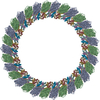
Journal: Structure / Year: 2023 Title: Structural basis of microtubule depolymerization by the kinesin-like activity of HIV-1 Rev. Authors: Elif Eren / Norman R Watts / Davide Randazzo / Ira Palmer / Dan L Sackett / Paul T Wingfield / Abstract: HIV-1 Rev is an essential regulatory protein that transports unspliced and partially spliced viral mRNAs from the nucleus to the cytoplasm for the expression of viral structural proteins. During its ...HIV-1 Rev is an essential regulatory protein that transports unspliced and partially spliced viral mRNAs from the nucleus to the cytoplasm for the expression of viral structural proteins. During its nucleocytoplasmic shuttling, Rev interacts with several host proteins to use the cellular machinery for the advantage of the virus. Here, we report the 3.5 Å cryo-EM structure of a 4.8 MDa Rev-tubulin ring complex. Our structure shows that Rev's arginine-rich motif (ARM) binds to both the acidic surfaces and the C-terminal tails of α/β-tubulin. The Rev-tubulin interaction is functionally homologous to that of kinesin-13, potently destabilizing microtubules at sub-stoichiometric levels. Expression of Rev in astrocytes and HeLa cells shows that it can modulate the microtubule cytoskeleton within the cellular environment. These results show a previously undefined regulatory role of Rev.
In the structure databanks used in Yorodumi, some data are registered as the other names, "COVID-19 virus" and "2019-nCoV". Here are the details of the virus and the list of structure data.
Jan 31, 2019. EMDB accession codes are about to change! (news from PDBe EMDB page)
EMDB accession codes are about to change! (news from PDBe EMDB page)
The allocation of 4 digits for EMDB accession codes will soon come to an end. Whilst these codes will remain in use, new EMDB accession codes will include an additional digit and will expand incrementally as the available range of codes is exhausted. The current 4-digit format prefixed with “EMD-” (i.e. EMD-XXXX) will advance to a 5-digit format (i.e. EMD-XXXXX), and so on. It is currently estimated that the 4-digit codes will be depleted around Spring 2019, at which point the 5-digit format will come into force.
The EM Navigator/Yorodumi systems omit the EMD- prefix.
Related info.:Q: What is EMD? / ID/Accession-code notation in Yorodumi/EM Navigator
Yorodumi is a browser for structure data from EMDB, PDB, SASBDB, etc.
This page is also the successor to EM Navigator detail page, and also detail information page/front-end page for Omokage search.
The word "yorodu" (or yorozu) is an old Japanese word meaning "ten thousand". "mi" (miru) is to see.
Related info.:EMDB / PDB / SASBDB / Comparison of 3 databanks / Yorodumi Search / Aug 31, 2016. New EM Navigator & Yorodumi / Yorodumi Papers / Jmol/JSmol / Function and homology information / Changes in new EM Navigator and Yorodumi
 Movie
Movie Controller


 Open data
 Basic information
 Components
 Keywords
 Function and homology information
 Human immunodeficiency virus 1
 Authors
 United States, 1items
  Citation
 Structure visualization
 Downloads & links
 Other downloads
 PDBj
PDBj


 Assembly
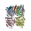


 Sample preparation
 Electron microscopy imaging
 FIELD EMISSION GUN / Accelerating voltage: 300 kV / Illumination mode: FLOOD BEAM
 Processing