 ムービー
ムービー コントローラー


+ データを開く

- 基本情報
基本情報
| 登録情報 | データベース: PDB / ID: 7u08 | |||||||||
|---|---|---|---|---|---|---|---|---|---|---|
| タイトル | Structure of CD148 fibronectin type III domain 1 and 2 | |||||||||
 要素 要素 | Receptor-type tyrosine-protein phosphatase eta | |||||||||
 キーワード キーワード | SIGNALING PROTEIN / receptor type protein tyrosine phosphatase | |||||||||
| 機能・相同性 |  機能・相同性情報 機能・相同性情報contact inhibition / positive regulation of Fc receptor mediated stimulatory signaling pathway / gamma-catenin binding / delta-catenin binding / positive regulation of platelet activation / negative regulation of vascular permeability / platelet-derived growth factor receptor binding / mitogen-activated protein kinase binding / negative regulation of platelet-derived growth factor receptor signaling pathway / platelet formation ...contact inhibition / positive regulation of Fc receptor mediated stimulatory signaling pathway / gamma-catenin binding / delta-catenin binding / positive regulation of platelet activation / negative regulation of vascular permeability / platelet-derived growth factor receptor binding / mitogen-activated protein kinase binding / negative regulation of platelet-derived growth factor receptor signaling pathway / platelet formation / negative regulation of T cell receptor signaling pathway / positive chemotaxis / positive regulation of macrophage chemotaxis / Phosphorylation of CD3 and TCR zeta chains / negative regulation of epidermal growth factor receptor signaling pathway / platelet-derived growth factor receptor signaling pathway / phosphatase activity / negative regulation of MAP kinase activity / peptidyl-tyrosine dephosphorylation / oligodendrocyte differentiation / immunological synapse / positive regulation of focal adhesion assembly / Negative regulation of FLT3 / vasculogenesis / regulation of cell adhesion / specific granule membrane / protein-tyrosine-phosphatase / negative regulation of phosphatidylinositol 3-kinase/protein kinase B signal transduction / positive regulation of cell adhesion / positive regulation of calcium-mediated signaling / negative regulation of insulin receptor signaling pathway / axon guidance / protein tyrosine phosphatase activity / positive regulation of phagocytosis / negative regulation of cell migration / B cell differentiation / Negative regulation of MET activity / negative regulation of cell growth / beta-catenin binding / ruffle membrane / cytokine-mediated signaling pathway / blood coagulation / positive regulation of tumor necrosis factor production / cell-cell junction / cell junction / glucose homeostasis / T cell receptor signaling pathway / heart development / angiogenesis / positive regulation of phosphatidylinositol 3-kinase/protein kinase B signal transduction / positive regulation of MAPK cascade / nuclear body / cadherin binding / negative regulation of cell population proliferation / Neutrophil degranulation / protein kinase binding / nucleolus / cell surface / signal transduction / extracellular exosome / nucleoplasm / plasma membrane 類似検索 - 分子機能 | |||||||||
| 生物種 |  Homo sapiens (ヒト) Homo sapiens (ヒト) | |||||||||
| 手法 |  X線回折 / シンクロトロン / 単波長異常分散 / 解像度: 3.30691735712 Å X線回折 / シンクロトロン / 単波長異常分散 / 解像度: 3.30691735712 Å | |||||||||
 データ登録者 データ登録者 | Zhou, D. / Zhu, J. | |||||||||
| 資金援助 |  米国, 2件 米国, 2件
| |||||||||
 引用 引用 | ジャーナル: To Be Published タイトル: Structure of CD148 fibronectin type III domain 1 and 2 著者: Zhou, D. / Zhu, J. | |||||||||
| 履歴 |
|
- 構造の表示
構造の表示
| 構造ビューア | 分子: MolmilJmol/JSmol |
|---|
- ダウンロードとリンク
-ダウンロード
| PDBx/mmCIF形式 | 7u08.cif.gz | 110.5 KB | 表示 | PDBx/mmCIF形式 |
|---|---|---|---|---|
| PDB形式 | pdb7u08.ent.gz | 71.1 KB | 表示 | PDB形式 |
| PDBx/mmJSON形式 | 7u08.json.gz | ツリー表示 | PDBx/mmJSON形式 | |
| その他 |  その他のダウンロード その他のダウンロード |
-検証レポート
| 文書・要旨 | 7u08_validation.pdf.gz | 428.1 KB | 表示 | wwPDB検証レポート |
|---|---|---|---|---|
| 文書・詳細版 | 7u08_full_validation.pdf.gz | 433 KB | 表示 | |
| XML形式データ | 7u08_validation.xml.gz | 9.7 KB | 表示 | |
| CIF形式データ | 7u08_validation.cif.gz | 11.7 KB | 表示 | |
| アーカイブディレクトリ | https://data.pdbj.org/pub/pdb/validation_reports/u0/7u08ftp://data.pdbj.org/pub/pdb/validation_reports/u0/7u08 | HTTPS FTP |
-関連構造データ

-リンク
 PDBj
PDBj


- 集合体
集合体
| 登録構造単位 | 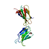
| ||||||||||||
|---|---|---|---|---|---|---|---|---|---|---|---|---|---|
| 1 |
| ||||||||||||
| 単位格子 |
|
-要素
| #1: タンパク質 | 分子量: 35729.773 Da / 分子数: 1 / 由来タイプ: 組換発現 / 由来: (組換発現) Homo sapiens (ヒト) / 遺伝子: PTPRJ, DEP1 / 発現宿主:  |
|---|---|
| #2: 化合物 | ChemComp-PT /   分子量: 195.078 Da / 分子数: 1 / 由来タイプ: 合成 / 式: Pt 分子量: 195.078 Da / 分子数: 1 / 由来タイプ: 合成 / 式: Pt |
| 研究の焦点であるリガンドがあるか | N |
| Has protein modification | Y |
-実験情報
-実験
| 実験 | 手法: X線回折 / 使用した結晶の数: 1 |
|---|
- 試料調製
試料調製
| 結晶 | マシュー密度: 2.36 Å3/Da / 溶媒含有率: 47.92 % |
|---|---|
| 結晶化 | 温度: 293 K / 手法: 蒸気拡散法, ハンギングドロップ法 / pH: 7.5 / 詳細: 0.25M MgCl2, 0.1M HEPES pH7.5, 25% PEG3350 |
-データ収集
| 回折 | 平均測定温度: 100 K / Serial crystal experiment: N |
|---|---|
| 放射光源 | 由来: シンクロトロン / サイト: APS / ビームライン: 21-ID-D / 波長: 0.9785 Å |
| 検出器 | タイプ: MARMOSAIC 300 mm CCD / 検出器: CCD / 日付: 2015年7月26日 |
| 放射 | プロトコル: SINGLE WAVELENGTH / 単色(M)・ラウエ(L): M / 散乱光タイプ: x-ray |
| 放射波長 | 波長: 0.9785 Å / 相対比: 1 |
| 反射 | 解像度: 3.3→20 Å / Num. obs: 5502 / % possible obs: 100 % / 冗長度: 38.8 % / Biso Wilson estimate: 135.736775868 Å2 / Rmerge(I) obs: 0.158 / Net I/σ(I): 34.3 |
| 反射 シェル | 解像度: 3.3→3.36 Å / Num. unique obs: 258 / CC1/2: 0.235 |
- 解析
解析
| ソフトウェア |
| ||||||||||||||||||||||||||||||||||||||||
|---|---|---|---|---|---|---|---|---|---|---|---|---|---|---|---|---|---|---|---|---|---|---|---|---|---|---|---|---|---|---|---|---|---|---|---|---|---|---|---|---|---|
| 精密化 | 構造決定の手法: 単波長異常分散 / 解像度: 3.30691735712→19.7369263569 Å / SU ML: 0.48564175729 / 交差検証法: FREE R-VALUE / σ(F): 0 / 位相誤差: 37.8756769017 立体化学のターゲット値: GeoStd + Monomer Library + CDL v1.2
| ||||||||||||||||||||||||||||||||||||||||
| 溶媒の処理 | 減衰半径: 0.9 Å / VDWプローブ半径: 1.11 Å / 溶媒モデル: FLAT BULK SOLVENT MODEL | ||||||||||||||||||||||||||||||||||||||||
| 原子変位パラメータ | Biso mean: 155.859958037 Å2 | ||||||||||||||||||||||||||||||||||||||||
| 精密化ステップ | サイクル: LAST / 解像度: 3.30691735712→19.7369263569 Å
| ||||||||||||||||||||||||||||||||||||||||
| 拘束条件 |
| ||||||||||||||||||||||||||||||||||||||||
| LS精密化 シェル |
| ||||||||||||||||||||||||||||||||||||||||
| 精密化 TLS | 手法: refined / Origin x: 30.1465290208 Å / Origin y: 40.7807218104 Å / Origin z: 31.6548398961 Å
| ||||||||||||||||||||||||||||||||||||||||
| 精密化 TLSグループ | Selection details: all |
