 Movie
Movie Controller


+ Open data

- Basic information
| Entry | Database: PDB / ID: 7u01 | |||||||||
|---|---|---|---|---|---|---|---|---|---|---|
| Title | Structure of CD148 fibronectin type III domain 2 | |||||||||
 Components Components | Receptor-type tyrosine-protein phosphatase eta | |||||||||
 Keywords Keywords | SIGNALING PROTEIN / receptor type protein tyrosine phosphatase | |||||||||
| Function / homology |  Function and homology information Function and homology informationcontact inhibition / positive regulation of Fc receptor mediated stimulatory signaling pathway / gamma-catenin binding / delta-catenin binding / positive regulation of platelet activation / negative regulation of vascular permeability / platelet-derived growth factor receptor binding / mitogen-activated protein kinase binding / platelet formation / negative regulation of platelet-derived growth factor receptor signaling pathway ...contact inhibition / positive regulation of Fc receptor mediated stimulatory signaling pathway / gamma-catenin binding / delta-catenin binding / positive regulation of platelet activation / negative regulation of vascular permeability / platelet-derived growth factor receptor binding / mitogen-activated protein kinase binding / platelet formation / negative regulation of platelet-derived growth factor receptor signaling pathway / negative regulation of T cell receptor signaling pathway / positive chemotaxis / Phosphorylation of CD3 and TCR zeta chains / positive regulation of macrophage chemotaxis / platelet-derived growth factor receptor signaling pathway / negative regulation of MAP kinase activity / peptidyl-tyrosine dephosphorylation / phosphatase activity / oligodendrocyte differentiation / negative regulation of epidermal growth factor receptor signaling pathway / immunological synapse / positive regulation of focal adhesion assembly / vasculogenesis / regulation of cell adhesion / specific granule membrane / protein-tyrosine-phosphatase / positive regulation of cell adhesion / Negative regulation of FLT3 / positive regulation of calcium-mediated signaling / negative regulation of insulin receptor signaling pathway / negative regulation of phosphatidylinositol 3-kinase/protein kinase B signal transduction / protein tyrosine phosphatase activity / axon guidance / B cell differentiation / negative regulation of cell migration / positive regulation of phagocytosis / Negative regulation of MET activity / negative regulation of cell growth / beta-catenin binding / ruffle membrane / cytokine-mediated signaling pathway / blood coagulation / positive regulation of tumor necrosis factor production / cell-cell junction / cell junction / T cell receptor signaling pathway / glucose homeostasis / heart development / angiogenesis / positive regulation of phosphatidylinositol 3-kinase/protein kinase B signal transduction / positive regulation of MAPK cascade / nuclear body / cadherin binding / negative regulation of cell population proliferation / Neutrophil degranulation / protein kinase binding / nucleolus / cell surface / signal transduction / extracellular exosome / nucleoplasm / plasma membrane Similarity search - Function | |||||||||
| Biological species |  Homo sapiens (human) Homo sapiens (human) | |||||||||
| Method |  X-RAY DIFFRACTION / SYNCHROTRON / SAD / Resolution: 2.29707910798 Å X-RAY DIFFRACTION / SYNCHROTRON / SAD / Resolution: 2.29707910798 Å | |||||||||
 Authors Authors | Zhou, D. / Zhu, J. | |||||||||
| Funding support |  United States, 2items United States, 2items
| |||||||||
 Citation Citation | Journal: To Be Published Title: Structure of CD148 fibronectin type III domain 1 and 2 Authors: Zhou, D. / Zhu, J. | |||||||||
| History |
|
- Structure visualization
| Structure viewer | Molecule: MolmilJmol/JSmol |
|---|
- Downloads & links
-Download
| PDBx/mmCIF format | 7u01.cif.gz | 211.1 KB | Display | PDBx/mmCIF format |
|---|---|---|---|---|
| PDB format | pdb7u01.ent.gz | 140.3 KB | Display | PDB format |
| PDBx/mmJSON format | 7u01.json.gz | Tree view | PDBx/mmJSON format | |
| Others |  Other downloads Other downloads |
-Validation report
| Arichive directory | https://data.pdbj.org/pub/pdb/validation_reports/u0/7u01ftp://data.pdbj.org/pub/pdb/validation_reports/u0/7u01 | HTTPS FTP |
|---|
-Related structure data

-Links
 PDBj
PDBj


- Assembly
| Deposited unit | 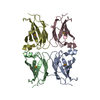
| ||||||||||||
|---|---|---|---|---|---|---|---|---|---|---|---|---|---|
| 1 | 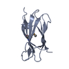
| ||||||||||||
| 2 | 
| ||||||||||||
| 3 | 
| ||||||||||||
| 4 | 
| ||||||||||||
| Unit cell |
| ||||||||||||
| Components on special symmetry positions |
|
-Components
| #1: Protein | Mass: 16958.318 Da / Num. of mol.: 4 Source method: isolated from a genetically manipulated source Source: (gene. exp.) Homo sapiens (human) / Gene: PTPRJ, DEP1 / Production host:  #2: Water | ChemComp-HOH / |  Mass: 18.015 Da / Num. of mol.: 91 / Source method: isolated from a natural source / Formula: H2O Mass: 18.015 Da / Num. of mol.: 91 / Source method: isolated from a natural source / Formula: H2OHas ligand of interest | Y | Has protein modification | Y | |
|---|
-Experimental details
-Experiment
| Experiment | Method: X-RAY DIFFRACTION / Number of used crystals: 1 |
|---|
- Sample preparation
| Crystal | Density Matthews: 2.13 Å3/Da / Density % sol: 42.29 % |
|---|---|
| Crystal grow | Temperature: 293 K / Method: vapor diffusion, hanging drop / pH: 4.5 / Details: 0.1M sodium acetate pH4.5, 3M NaCl |
-Data collection
| Diffraction | Mean temperature: 100 K / Serial crystal experiment: N |
|---|---|
| Diffraction source | Source: SYNCHROTRON / Site: APS / Beamline: 21-ID-D / Wavelength: 0.979276 Å |
| Detector | Type: MARMOSAIC 300 mm CCD / Detector: CCD / Date: Jun 7, 2016 |
| Radiation | Protocol: SINGLE WAVELENGTH / Monochromatic (M) / Laue (L): M / Scattering type: x-ray |
| Radiation wavelength | Wavelength: 0.979276 Å / Relative weight: 1 |
| Reflection | Resolution: 2.297→50 Å / Num. obs: 26929 / % possible obs: 100 % / Redundancy: 26.6 % / Biso Wilson estimate: 54.4779195349 Å2 / Rmerge(I) obs: 0.098 / Net I/σ(I): 22.4 |
| Reflection shell | Resolution: 2.2971→2.3545 Å / Rmerge(I) obs: 1.1 / Num. unique obs: 1712 |
- Processing
| Software |
| |||||||||||||||||||||||||||||||||||||||||||||||||||||||||||||||||||||||||||||||||||||||||||||||||||||||||
|---|---|---|---|---|---|---|---|---|---|---|---|---|---|---|---|---|---|---|---|---|---|---|---|---|---|---|---|---|---|---|---|---|---|---|---|---|---|---|---|---|---|---|---|---|---|---|---|---|---|---|---|---|---|---|---|---|---|---|---|---|---|---|---|---|---|---|---|---|---|---|---|---|---|---|---|---|---|---|---|---|---|---|---|---|---|---|---|---|---|---|---|---|---|---|---|---|---|---|---|---|---|---|---|---|---|---|
| Refinement | Method to determine structure: SAD / Resolution: 2.29707910798→45.7113330479 Å / SU ML: 0.329266292209 / Cross valid method: FREE R-VALUE / σ(F): 1.35298695758 / Phase error: 31.7182115406 Stereochemistry target values: GeoStd + Monomer Library + CDL v1.2
| |||||||||||||||||||||||||||||||||||||||||||||||||||||||||||||||||||||||||||||||||||||||||||||||||||||||||
| Solvent computation | Shrinkage radii: 0.9 Å / VDW probe radii: 1.11 Å / Solvent model: FLAT BULK SOLVENT MODEL | |||||||||||||||||||||||||||||||||||||||||||||||||||||||||||||||||||||||||||||||||||||||||||||||||||||||||
| Displacement parameters | Biso mean: 69.786072096 Å2 | |||||||||||||||||||||||||||||||||||||||||||||||||||||||||||||||||||||||||||||||||||||||||||||||||||||||||
| Refinement step | Cycle: LAST / Resolution: 2.29707910798→45.7113330479 Å
| |||||||||||||||||||||||||||||||||||||||||||||||||||||||||||||||||||||||||||||||||||||||||||||||||||||||||
| Refine LS restraints |
| |||||||||||||||||||||||||||||||||||||||||||||||||||||||||||||||||||||||||||||||||||||||||||||||||||||||||
| LS refinement shell |
| |||||||||||||||||||||||||||||||||||||||||||||||||||||||||||||||||||||||||||||||||||||||||||||||||||||||||
| Refinement TLS params. | Method: refined / Origin x: 116.185225815 Å / Origin y: 76.2849144805 Å / Origin z: 19.365837285 Å
| |||||||||||||||||||||||||||||||||||||||||||||||||||||||||||||||||||||||||||||||||||||||||||||||||||||||||
| Refinement TLS group | Selection details: all |
