 Movie
Movie Controller

[English] 日本語
 Yorodumi
- PDB-7tym: Cryo-EM Structure of insulin receptor-related receptor (IRR) in a... -
+ Open data

- Basic information
| Entry | Database: PDB / ID: 7tym | |||||||||
|---|---|---|---|---|---|---|---|---|---|---|
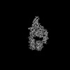
| Title | Cryo-EM Structure of insulin receptor-related receptor (IRR) in active-state captured at pH 9. The 3D refinement was applied with C2 symmetry | |||||||||
 Components Components | Insulin receptor-related protein | |||||||||
 Keywords Keywords | SIGNALING PROTEIN / Receptor tyrosine kinase / insulin receptor family | |||||||||
| Function / homology |  Function and homology information Function and homology informationcellular response to alkaline pH / male sex determination / insulin receptor complex / insulin receptor activity / insulin receptor substrate binding / phosphatidylinositol 3-kinase binding / transmembrane receptor protein tyrosine kinase activity / cell surface receptor protein tyrosine kinase signaling pathway / receptor protein-tyrosine kinase / insulin receptor signaling pathway ...cellular response to alkaline pH / male sex determination / insulin receptor complex / insulin receptor activity / insulin receptor substrate binding / phosphatidylinositol 3-kinase binding / transmembrane receptor protein tyrosine kinase activity / cell surface receptor protein tyrosine kinase signaling pathway / receptor protein-tyrosine kinase / insulin receptor signaling pathway / protein autophosphorylation / actin cytoskeleton organization / receptor complex / axon / ATP binding / plasma membrane Similarity search - Function | |||||||||
| Biological species |  Homo sapiens (human) Homo sapiens (human) | |||||||||
| Method | ELECTRON MICROSCOPY / single particle reconstruction / cryo EM / Resolution: 3.4 Å | |||||||||
 Authors Authors | Wang, L.W. / Hall, C. / Li, J. / Choi, E. / Bai, X.C. | |||||||||
| Funding support |  United States, 2items United States, 2items
| |||||||||
 Citation Citation | Journal: Nat Struct Mol Biol / Year: 2023 Title: Structural basis of the alkaline pH-dependent activation of insulin receptor-related receptor. Authors: Liwei Wang / Catherine Hall / Jie Li / Eunhee Choi / Xiao-Chen Bai / Abstract: The insulin receptor (IR) family is a subfamily of receptor tyrosine kinases that controls metabolic homeostasis and cell growth. Distinct from IR and insulin-like growth factor 1 receptor, whose ...The insulin receptor (IR) family is a subfamily of receptor tyrosine kinases that controls metabolic homeostasis and cell growth. Distinct from IR and insulin-like growth factor 1 receptor, whose activation requires ligand binding, insulin receptor-related receptor (IRR)-the third member of the IR family-is activated by alkaline pH. However, the molecular mechanism underlying alkaline pH-induced IRR activation remains unclear. Here, we present cryo-EM structures of human IRR in both neutral pH inactive and alkaline pH active states. Combined with mutagenesis and cellular assays, we show that, upon pH increase, electrostatic repulsion of the pH-sensitive motifs of IRR disrupts its autoinhibited state and promotes a scissor-like rotation between two protomers, leading to a T-shaped active conformation. Together, our study reveals an unprecedented alkaline pH-dependent activation mechanism of IRR, opening up opportunities to understand the structure-function relationship of this important receptor. | |||||||||
| History |
|
- Structure visualization
| Structure viewer | Molecule: MolmilJmol/JSmol |
|---|
- Downloads & links
-Download
| PDBx/mmCIF format | 7tym.cif.gz | 329.8 KB | Display | PDBx/mmCIF format |
|---|---|---|---|---|
| PDB format | pdb7tym.ent.gz | 251.4 KB | Display | PDB format |
| PDBx/mmJSON format | 7tym.json.gz | Tree view | PDBx/mmJSON format | |
| Others |  Other downloads Other downloads |
-Validation report
| Summary document | 7tym_validation.pdf.gz | 890.6 KB | Display | wwPDB validaton report |
|---|---|---|---|---|
| Full document | 7tym_full_validation.pdf.gz | 906.2 KB | Display | |
| Data in XML | 7tym_validation.xml.gz | 44.9 KB | Display | |
| Data in CIF | 7tym_validation.cif.gz | 66.9 KB | Display | |
| Arichive directory | https://data.pdbj.org/pub/pdb/validation_reports/ty/7tymftp://data.pdbj.org/pub/pdb/validation_reports/ty/7tym | HTTPS FTP |
-Related structure data
| Related structure data |  26185MC  7tyjC  7tykC C: citing same article ( M: map data used to model this data |
|---|---|
| Similar structure data |

-Links
 PDBj
PDBj


- Assembly
| Deposited unit | 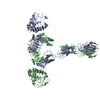
|
|---|---|
| 1 |
|
-Components
| #1: Protein | Mass: 143879.547 Da / Num. of mol.: 2 Source method: isolated from a genetically manipulated source Source: (gene. exp.) Homo sapiens (human) / Gene: INSRR, IRR / Production host: Homo sapiens (human)References: UniProt: P14616, receptor protein-tyrosine kinase Has protein modification | Y | |
|---|
-Experimental details
-Experiment
| Experiment | Method: ELECTRON MICROSCOPY |
|---|---|
| EM experiment | Aggregation state: PARTICLE / 3D reconstruction method: single particle reconstruction |
- Sample preparation
| Component | Name: Insulin receptor-related receptor (IRR) in active-state captured at pH 9 Type: COMPLEX / Entity ID: all / Source: RECOMBINANT |
|---|---|
| Molecular weight | Value: 0.14 MDa / Experimental value: NO |
| Source (natural) | Organism: Homo sapiens (human) |
| Source (recombinant) | Organism: Homo sapiens (human) |
| Buffer solution | pH: 9 |
| Specimen | Conc.: 6 mg/ml / Embedding applied: NO / Shadowing applied: NO / Staining applied: NO / Vitrification applied: YES |
| Vitrification | Cryogen name: ETHANE / Humidity: 100 % |
- Electron microscopy imaging
| Experimental equipment |  Model: Titan Krios / Image courtesy: FEI Company |
|---|---|
| Microscopy | Model: FEI TITAN KRIOS |
| Electron gun | Electron source:  FIELD EMISSION GUN / Accelerating voltage: 300 kV / Illumination mode: FLOOD BEAM FIELD EMISSION GUN / Accelerating voltage: 300 kV / Illumination mode: FLOOD BEAM |
| Electron lens | Mode: BRIGHT FIELD / Nominal defocus max: 2600 nm / Nominal defocus min: 1600 nm |
| Specimen holder | Cryogen: NITROGEN / Specimen holder model: FEI TITAN KRIOS AUTOGRID HOLDER |
| Image recording | Electron dose: 60 e/Å2 / Film or detector model: GATAN K3 BIOQUANTUM (6k x 4k) |
| EM imaging optics | Energyfilter name: GIF Bioquantum / Energyfilter slit width: 20 eV |
- Processing
| EM software |
| |||||||||||||||||||||||||||
|---|---|---|---|---|---|---|---|---|---|---|---|---|---|---|---|---|---|---|---|---|---|---|---|---|---|---|---|---|
| CTF correction | Type: PHASE FLIPPING AND AMPLITUDE CORRECTION | |||||||||||||||||||||||||||
| Particle selection | Num. of particles selected: 3034847 | |||||||||||||||||||||||||||
| 3D reconstruction | Resolution: 3.4 Å / Resolution method: FSC 0.143 CUT-OFF / Num. of particles: 79010 / Symmetry type: POINT | |||||||||||||||||||||||||||
| Atomic model building | Protocol: AB INITIO MODEL / Space: REAL |
