 Movie
Movie Controller

[English] 日本語
 Yorodumi
- PDB-7tyd: Crystal structure of FGFR4 domain 3 in complex with a de novo-des... -
+ Open data

- Basic information
| Entry | Database: PDB / ID: 7tyd | ||||||
|---|---|---|---|---|---|---|---|
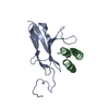
| Title | Crystal structure of FGFR4 domain 3 in complex with a de novo-designed mini-binder in P21 space group | ||||||
 Components Components |
| ||||||
 Keywords Keywords | SIGNALING PROTEIN / Receptor tyrosine kinase / Complex / Binder / MEMBRANE PROTEIN | ||||||
| Function / homology |  Function and homology information Function and homology informationFGFR4 mutant receptor activation / betaKlotho-mediated ligand binding / regulation of extracellular matrix disassembly / positive regulation of catalytic activity / phosphate ion homeostasis / regulation of bile acid biosynthetic process / FGFR4 ligand binding and activation / Phospholipase C-mediated cascade; FGFR4 / fibroblast growth factor receptor activity / positive regulation of DNA biosynthetic process ...FGFR4 mutant receptor activation / betaKlotho-mediated ligand binding / regulation of extracellular matrix disassembly / positive regulation of catalytic activity / phosphate ion homeostasis / regulation of bile acid biosynthetic process / FGFR4 ligand binding and activation / Phospholipase C-mediated cascade; FGFR4 / fibroblast growth factor receptor activity / positive regulation of DNA biosynthetic process / PI-3K cascade:FGFR4 / fibroblast growth factor binding / positive regulation of proteolysis / regulation of lipid metabolic process / PI3K Cascade / fibroblast growth factor receptor signaling pathway / SHC-mediated cascade:FGFR4 / transport vesicle / Signaling by FGFR4 in disease / FRS-mediated FGFR4 signaling / peptidyl-tyrosine phosphorylation / cholesterol homeostasis / Negative regulation of FGFR4 signaling / receptor protein-tyrosine kinase / Constitutive Signaling by Aberrant PI3K in Cancer / cell migration / PIP3 activates AKT signaling / glucose homeostasis / heparin binding / protein autophosphorylation / PI5P, PP2A and IER3 Regulate PI3K/AKT Signaling / RAF/MAP kinase cascade / positive regulation of ERK1 and ERK2 cascade / receptor complex / endosome / positive regulation of cell population proliferation / positive regulation of gene expression / endoplasmic reticulum / Golgi apparatus / extracellular region / ATP binding / plasma membrane Similarity search - Function | ||||||
| Biological species |  Homo sapiens (human) Homo sapiens (human)synthetic construct (others) | ||||||
| Method |  X-RAY DIFFRACTION / SYNCHROTRON / MOLECULAR REPLACEMENT / Resolution: 2.86 Å X-RAY DIFFRACTION / SYNCHROTRON / MOLECULAR REPLACEMENT / Resolution: 2.86 Å | ||||||
 Authors Authors | Park, J.S. / Lee, S. | ||||||
| Funding support | 1items
| ||||||
 Citation Citation | Journal: Cell Rep / Year: 2022 Title: Isoform-specific inhibition of FGFR signaling achieved by a de-novo-designed mini-protein. Authors: Park, J.S. / Choi, J. / Cao, L. / Mohanty, J. / Suzuki, Y. / Park, A. / Baker, D. / Schlessinger, J. / Lee, S. | ||||||
| History |
|
- Structure visualization
| Structure viewer | Molecule: MolmilJmol/JSmol |
|---|
- Downloads & links
-Download
| PDBx/mmCIF format | 7tyd.cif.gz | 85.1 KB | Display | PDBx/mmCIF format |
|---|---|---|---|---|
| PDB format | pdb7tyd.ent.gz | 49.3 KB | Display | PDB format |
| PDBx/mmJSON format | 7tyd.json.gz | Tree view | PDBx/mmJSON format | |
| Others |  Other downloads Other downloads |
-Validation report
| Arichive directory | https://data.pdbj.org/pub/pdb/validation_reports/ty/7tydftp://data.pdbj.org/pub/pdb/validation_reports/ty/7tyd | HTTPS FTP |
|---|
-Related structure data
| Related structure data | 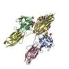 1cvsS S: Starting model for refinement |
|---|---|
| Similar structure data |
-Links
 PDBj
PDBj


- Assembly
| Deposited unit | 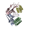
| ||||||||||||
|---|---|---|---|---|---|---|---|---|---|---|---|---|---|
| 1 | 
| ||||||||||||
| 2 | 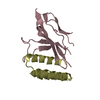
| ||||||||||||
| Unit cell |
|
-Components
| #1: Protein | Mass: 14271.884 Da / Num. of mol.: 2 Source method: isolated from a genetically manipulated source Source: (gene. exp.) Homo sapiens (human) / Gene: FGFR4, JTK2, TKF / Production host:  References: UniProt: P22455, receptor protein-tyrosine kinase #2: Protein | Mass: 7535.809 Da / Num. of mol.: 2 Source method: isolated from a genetically manipulated source Source: (gene. exp.) synthetic construct (others) / Production host: #3: Water | ChemComp-HOH / |  Mass: 18.015 Da / Num. of mol.: 6 / Source method: isolated from a natural source / Formula: H2O Mass: 18.015 Da / Num. of mol.: 6 / Source method: isolated from a natural source / Formula: H2OHas protein modification | Y | |
|---|
-Experimental details
-Experiment
| Experiment | Method: X-RAY DIFFRACTION / Number of used crystals: 1 |
|---|
- Sample preparation
| Crystal | Density Matthews: 2.26 Å3/Da / Density % sol: 45.5 % |
|---|---|
| Crystal grow | Temperature: 277 K / Method: vapor diffusion, hanging drop / pH: 5.5 Details: 0.1 M Bis-Tris (pH 5.5) 25% polyethylene glycol 3,350 3% 1,5-diaminopentane dihydrochloride |
-Data collection
| Diffraction | Mean temperature: 100 K / Serial crystal experiment: N |
|---|---|
| Diffraction source | Source: SYNCHROTRON / Site: APS  / Beamline: 24-ID-E / Wavelength: 0.97918 Å / Beamline: 24-ID-E / Wavelength: 0.97918 Å |
| Detector | Type: DECTRIS EIGER X 16M / Detector: PIXEL / Date: Mar 10, 2021 |
| Radiation | Protocol: SINGLE WAVELENGTH / Monochromatic (M) / Laue (L): M / Scattering type: x-ray |
| Radiation wavelength | Wavelength: 0.97918 Å / Relative weight: 1 |
| Reflection | Resolution: 2.85→50 Å / Num. obs: 8780 / % possible obs: 96.1 % / Redundancy: 4.9 % / CC1/2: 0.992 / Net I/σ(I): 18.55 |
| Reflection shell | Resolution: 2.85→2.9 Å / Redundancy: 4.1 % / Mean I/σ(I) obs: 2.564 / Num. unique obs: 412 / CC1/2: 0.889 / % possible all: 87.3 |
- Processing
| Software |
| ||||||||||||||||||||||||||||
|---|---|---|---|---|---|---|---|---|---|---|---|---|---|---|---|---|---|---|---|---|---|---|---|---|---|---|---|---|---|
| Refinement | Method to determine structure: MOLECULAR REPLACEMENT Starting model: 1CVS Resolution: 2.86→49.32 Å / SU ML: 0.4751 / Cross valid method: FREE R-VALUE / σ(F): 1.41 / Phase error: 37.347 Stereochemistry target values: GeoStd + Monomer Library + CDL v1.2
| ||||||||||||||||||||||||||||
| Solvent computation | Shrinkage radii: 0.9 Å / VDW probe radii: 1.11 Å / Solvent model: FLAT BULK SOLVENT MODEL | ||||||||||||||||||||||||||||
| Displacement parameters | Biso mean: 58.09 Å2 | ||||||||||||||||||||||||||||
| Refinement step | Cycle: LAST / Resolution: 2.86→49.32 Å
| ||||||||||||||||||||||||||||
| Refine LS restraints |
| ||||||||||||||||||||||||||||
| LS refinement shell |
|
