 Movie
Movie Controller

[English] 日本語
 Yorodumi
- PDB-7tyb: Salicylate Adenylate PchD from Pseudomonas aeruginosa containing ... -
+ Open data

- Basic information
| Entry | Database: PDB / ID: 7tyb | ||||||||||||
|---|---|---|---|---|---|---|---|---|---|---|---|---|---|
| Title | Salicylate Adenylate PchD from Pseudomonas aeruginosa containing salicyl-AMS | ||||||||||||
 Components Components | Pyochelin biosynthesis protein PchD | ||||||||||||
 Keywords Keywords | BIOSYNTHETIC PROTEIN / siderophore | ||||||||||||
| Function / homology |  Function and homology information Function and homology informationsalicylate-[aryl-carrier protein] ligase / ligase activity / antibiotic biosynthetic process Similarity search - Function | ||||||||||||
| Biological species |  Pseudomonas aeruginosa PAO1 (bacteria) Pseudomonas aeruginosa PAO1 (bacteria) | ||||||||||||
| Method |  X-RAY DIFFRACTION / SYNCHROTRON / MOLECULAR REPLACEMENT / Resolution: 2.11 Å X-RAY DIFFRACTION / SYNCHROTRON / MOLECULAR REPLACEMENT / Resolution: 2.11 Å | ||||||||||||
 Authors Authors | Meneely, K.M. / Shelton, C.L. / Lamb, A.L. | ||||||||||||
| Funding support |  United States, 3items United States, 3items
| ||||||||||||
 Citation Citation | Journal: J.Biol.Inorg.Chem. / Year: 2022 Title: Rational inhibitor design for Pseudomonas aeruginosa salicylate adenylation enzyme PchD. Authors: Shelton, C.L. / Meneely, K.M. / Ronnebaum, T.A. / Chilton, A.S. / Riley, A.P. / Prisinzano, T.E. / Lamb, A.L. | ||||||||||||
| History |
|
- Structure visualization
| Structure viewer | Molecule: MolmilJmol/JSmol |
|---|
- Downloads & links
-Download
| PDBx/mmCIF format | 7tyb.cif.gz | 251.9 KB | Display | PDBx/mmCIF format |
|---|---|---|---|---|
| PDB format | pdb7tyb.ent.gz | 164.9 KB | Display | PDB format |
| PDBx/mmJSON format | 7tyb.json.gz | Tree view | PDBx/mmJSON format | |
| Others |  Other downloads Other downloads |
-Validation report
| Arichive directory | https://data.pdbj.org/pub/pdb/validation_reports/ty/7tybftp://data.pdbj.org/pub/pdb/validation_reports/ty/7tyb | HTTPS FTP |
|---|
-Related structure data
| Related structure data | 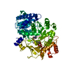 7tz4C  1mdbS S: Starting model for refinement C: citing same article ( |
|---|---|
| Similar structure data |
-Links
 PDBj
PDBj
- Assembly
| Deposited unit | 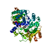
| ||||||||||||
|---|---|---|---|---|---|---|---|---|---|---|---|---|---|
| 1 |
| ||||||||||||
| Unit cell |
|
-Components
| #1: Protein | Mass: 59982.332 Da / Num. of mol.: 1 Source method: isolated from a genetically manipulated source Source: (gene. exp.) Pseudomonas aeruginosa PAO1 (bacteria)Strain: ATCC 15692 / DSM 22644 / CIP 104116 / JCM 14847 / LMG 12228 / 1C / PRS 101 / PAO1 Gene: pchD, PA4228 / Production host: |
|---|---|
| #2: Chemical | ChemComp-KT0 /   Mass: 466.425 Da / Num. of mol.: 1 / Source method: obtained synthetically / Formula: C17H18N6O8S / Feature type: SUBJECT OF INVESTIGATION Mass: 466.425 Da / Num. of mol.: 1 / Source method: obtained synthetically / Formula: C17H18N6O8S / Feature type: SUBJECT OF INVESTIGATION |
| #3: Water | ChemComp-HOH /  Mass: 18.015 Da / Num. of mol.: 139 / Source method: isolated from a natural source / Formula: H2O Mass: 18.015 Da / Num. of mol.: 139 / Source method: isolated from a natural source / Formula: H2O |
| Has ligand of interest | Y |
-Experimental details
-Experiment
| Experiment | Method: X-RAY DIFFRACTION / Number of used crystals: 1 |
|---|
- Sample preparation
| Crystal | Density Matthews: 2.2 Å3/Da / Density % sol: 43.98 % |
|---|---|
| Crystal grow | Temperature: 298 K / Method: vapor diffusion, hanging drop / pH: 5.4 Details: 0.2 M ammonium acetate, 0.1 M MES pH 5.4, 0.03 M ammonium chloride, 20% PEG 8000 |
-Data collection
| Diffraction | Mean temperature: 100 K / Serial crystal experiment: N |
|---|---|
| Diffraction source | Source: SYNCHROTRON / Site: SSRL / Beamline: BL7-1 / Wavelength: 1.1271 Å |
| Detector | Type: ADSC QUANTUM 315r / Detector: CCD / Date: Jan 23, 2016 / Details: Mirror: Rh coated flat bent mirror |
| Radiation | Monochromator: Si(111) side scattering I-beam bent single crystal; asymmetric cut 4.9650 deg Protocol: SINGLE WAVELENGTH / Monochromatic (M) / Laue (L): M / Scattering type: x-ray |
| Radiation wavelength | Wavelength: 1.1271 Å / Relative weight: 1 |
| Reflection | Resolution: 2.11→39.48 Å / Num. obs: 29938 / % possible obs: 99.1 % / Redundancy: 5.5 % / Biso Wilson estimate: 17.06 Å2 / Rpim(I) all: 0.091 / Net I/σ(I): 7.5 |
| Reflection shell | Resolution: 2.11→2.18 Å / Redundancy: 5.1 % / Mean I/σ(I) obs: 2.2 / Num. unique obs: 2235 / Rpim(I) all: 0.426 / % possible all: 90.5 |
- Processing
| Software |
| ||||||||||||||||||||||||||||||||||||||||||||||||||||||||||||||||||||||||||||||||||||||||||||||||||
|---|---|---|---|---|---|---|---|---|---|---|---|---|---|---|---|---|---|---|---|---|---|---|---|---|---|---|---|---|---|---|---|---|---|---|---|---|---|---|---|---|---|---|---|---|---|---|---|---|---|---|---|---|---|---|---|---|---|---|---|---|---|---|---|---|---|---|---|---|---|---|---|---|---|---|---|---|---|---|---|---|---|---|---|---|---|---|---|---|---|---|---|---|---|---|---|---|---|---|---|
| Refinement | Method to determine structure: MOLECULAR REPLACEMENT Starting model: 1MDB Resolution: 2.11→37.01 Å / SU ML: 0.2384 / Cross valid method: FREE R-VALUE / σ(F): 0 / Phase error: 22.5111 Stereochemistry target values: GeoStd + Monomer Library + CDL v1.2
| ||||||||||||||||||||||||||||||||||||||||||||||||||||||||||||||||||||||||||||||||||||||||||||||||||
| Solvent computation | Shrinkage radii: 0.9 Å / VDW probe radii: 1.11 Å / Solvent model: FLAT BULK SOLVENT MODEL | ||||||||||||||||||||||||||||||||||||||||||||||||||||||||||||||||||||||||||||||||||||||||||||||||||
| Displacement parameters | Biso mean: 23.99 Å2 | ||||||||||||||||||||||||||||||||||||||||||||||||||||||||||||||||||||||||||||||||||||||||||||||||||
| Refinement step | Cycle: LAST / Resolution: 2.11→37.01 Å
| ||||||||||||||||||||||||||||||||||||||||||||||||||||||||||||||||||||||||||||||||||||||||||||||||||
| Refine LS restraints |
| ||||||||||||||||||||||||||||||||||||||||||||||||||||||||||||||||||||||||||||||||||||||||||||||||||
| LS refinement shell |
|
