 Movie
Movie Controller

[English] 日本語
 Yorodumi
- PDB-7txp: X-ray structure of the VioB N-acetyltransferase from Acinetobacte... -
+ Open data

- Basic information
| Entry | Database: PDB / ID: 7txp | ||||||
|---|---|---|---|---|---|---|---|
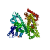
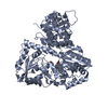
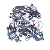
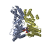
| Title | X-ray structure of the VioB N-acetyltransferase from Acinetobacter baumannii in complex with TDP-4-amino-4,6-dideoxy-D-glucose | ||||||
 Components Components | VioB | ||||||
 Keywords Keywords | TRANSFERASE / N-acetyltransferase / beta helix / 4-amino-4 / 6-dideoxy-D-glucose | ||||||
| Function / homology |  Function and homology information Function and homology information | ||||||
| Biological species |  Acinetobacter baumannii (bacteria) Acinetobacter baumannii (bacteria) | ||||||
| Method |  X-RAY DIFFRACTION / MOLECULAR REPLACEMENT / Resolution: 1.45 Å X-RAY DIFFRACTION / MOLECULAR REPLACEMENT / Resolution: 1.45 Å | ||||||
 Authors Authors | Herkert, N.R. / Thoden, J.B. / Holden, H.M. | ||||||
| Funding support |  United States, 1items United States, 1items
| ||||||
 Citation Citation | Journal: Proteins / Year: 2022 Title: Structure and function of an N-acetyltransferase from the human pathogen Acinetobacter baumannii isolate BAL_212. Authors: Herkert, N.R. / Thoden, J.B. / Holden, H.M. | ||||||
| History |
|
- Structure visualization
| Structure viewer | Molecule: MolmilJmol/JSmol |
|---|
- Downloads & links
-Download
| PDBx/mmCIF format | 7txp.cif.gz | 63.1 KB | Display | PDBx/mmCIF format |
|---|---|---|---|---|
| PDB format | pdb7txp.ent.gz | 43.2 KB | Display | PDB format |
| PDBx/mmJSON format | 7txp.json.gz | Tree view | PDBx/mmJSON format | |
| Others |  Other downloads Other downloads |
-Validation report
| Arichive directory | https://data.pdbj.org/pub/pdb/validation_reports/tx/7txpftp://data.pdbj.org/pub/pdb/validation_reports/tx/7txp | HTTPS FTP |
|---|
-Related structure data
| Related structure data | 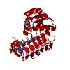 7txqC  7txsC  4m98S S: Starting model for refinement C: citing same article ( |
|---|---|
| Similar structure data |


-Links
 PDBj
PDBj


- Assembly
| Deposited unit | 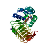
| ||||||||||||
|---|---|---|---|---|---|---|---|---|---|---|---|---|---|
| 1 | 
| ||||||||||||
| Unit cell |
| ||||||||||||
| Components on special symmetry positions |
|
-Components
| #1: Protein | Mass: 23305.889 Da / Num. of mol.: 1 Source method: isolated from a genetically manipulated source Source: (gene. exp.) Acinetobacter baumannii (bacteria) / Gene: vioB / Production host: | ||||
|---|---|---|---|---|---|
| #2: Chemical | ChemComp-0FX /   Mass: 547.345 Da / Num. of mol.: 1 / Source method: obtained synthetically / Formula: C16H27N3O14P2 / Feature type: SUBJECT OF INVESTIGATION Mass: 547.345 Da / Num. of mol.: 1 / Source method: obtained synthetically / Formula: C16H27N3O14P2 / Feature type: SUBJECT OF INVESTIGATION | ||||
| #3: Chemical |   Mass: 22.990 Da / Num. of mol.: 2 / Source method: obtained synthetically / Formula: Na Mass: 22.990 Da / Num. of mol.: 2 / Source method: obtained synthetically / Formula: Na#4: Water | ChemComp-HOH / |  Mass: 18.015 Da / Num. of mol.: 211 / Source method: isolated from a natural source / Formula: H2O Mass: 18.015 Da / Num. of mol.: 211 / Source method: isolated from a natural source / Formula: H2OHas ligand of interest | Y | |
-Experimental details
-Experiment
| Experiment | Method: X-RAY DIFFRACTION / Number of used crystals: 1 |
|---|
- Sample preparation
| Crystal | Density Matthews: 2.85 Å3/Da / Density % sol: 56.8 % |
|---|---|
| Crystal grow | Temperature: 293 K / Method: vapor diffusion, hanging drop / pH: 6 Details: 8 - 12% poly(ethylene glycol) 8000, 200 mM LiCl, 5 mM dTDP-4-amino-4,6-dideoxy-D-glucose, 5 mM CoA, and 100 mM MES (pH 6.0). Soak in ligand fre solution followed by soak in 20 mM dTDP-4- ...Details: 8 - 12% poly(ethylene glycol) 8000, 200 mM LiCl, 5 mM dTDP-4-amino-4,6-dideoxy-D-glucose, 5 mM CoA, and 100 mM MES (pH 6.0). Soak in ligand fre solution followed by soak in 20 mM dTDP-4-amino-4,6-dideoxy-D-glucose |
-Data collection
| Diffraction | Mean temperature: 100 K / Serial crystal experiment: N |
|---|---|
| Diffraction source | Source: SEALED TUBE / Type: BRUKER D8 QUEST / Wavelength: 1.5418 Å |
| Detector | Type: Bruker PHOTON II / Detector: PIXEL / Date: Aug 20, 2021 |
| Radiation | Protocol: SINGLE WAVELENGTH / Monochromatic (M) / Laue (L): M / Scattering type: x-ray |
| Radiation wavelength | Wavelength: 1.5418 Å / Relative weight: 1 |
| Reflection | Resolution: 1.45→50 Å / Num. obs: 45094 / % possible obs: 98.7 % / Observed criterion σ(F): 0 / Observed criterion σ(I): 0 / Redundancy: 4.2 % / Rsym value: 0.069 / Net I/σ(I): 10.4 |
| Reflection shell | Resolution: 1.45→1.55 Å / Redundancy: 2.9 % / Mean I/σ(I) obs: 3 / Num. unique obs: 8089 / Rsym value: 0.353 / % possible all: 96.8 |
- Processing
| Software |
| ||||||||||||||||||||||||||||||||||||||||||||||||||||||||||||
|---|---|---|---|---|---|---|---|---|---|---|---|---|---|---|---|---|---|---|---|---|---|---|---|---|---|---|---|---|---|---|---|---|---|---|---|---|---|---|---|---|---|---|---|---|---|---|---|---|---|---|---|---|---|---|---|---|---|---|---|---|---|
| Refinement | Method to determine structure: MOLECULAR REPLACEMENT Starting model: 4m98 Resolution: 1.45→36.47 Å / Cor.coef. Fo:Fc: 0.952 / Cor.coef. Fo:Fc free: 0.941 / SU B: 1.117 / SU ML: 0.043 / Cross valid method: THROUGHOUT / σ(F): 0 / ESU R: 0.059 / ESU R Free: 0.059 / Stereochemistry target values: MAXIMUM LIKELIHOOD Details: HYDROGENS HAVE BEEN ADDED IN THE RIDING POSITIONS U VALUES : REFINED INDIVIDUALLY
| ||||||||||||||||||||||||||||||||||||||||||||||||||||||||||||
| Solvent computation | Ion probe radii: 0.8 Å / Shrinkage radii: 0.8 Å / VDW probe radii: 1.2 Å / Solvent model: MASK | ||||||||||||||||||||||||||||||||||||||||||||||||||||||||||||
| Displacement parameters | Biso max: 48.03 Å2 / Biso mean: 12.155 Å2 / Biso min: 4.53 Å2
| ||||||||||||||||||||||||||||||||||||||||||||||||||||||||||||
| Refinement step | Cycle: final / Resolution: 1.45→36.47 Å
| ||||||||||||||||||||||||||||||||||||||||||||||||||||||||||||
| Refine LS restraints |
| ||||||||||||||||||||||||||||||||||||||||||||||||||||||||||||
| LS refinement shell | Resolution: 1.45→1.487 Å / Rfactor Rfree error: 0
|
