 Movie
Movie Controller

[English] 日本語
 Yorodumi
- PDB-7tx2: Crystal structure of human phenylethanolamine N-methyltransferase... -
+ Open data

- Basic information
| Entry | Database: PDB / ID: 7tx2 | ||||||
|---|---|---|---|---|---|---|---|
| Title | Crystal structure of human phenylethanolamine N-methyltransferase (PNMT) in complex with (2S)-2-amino-4-(((5-(6-amino-9H-purin-9-yl)-3,4-dihydroxytetrahydrofuran-2-yl)methyl)(4-(7,8-dichloro-1,2,3,4-tetrahydroisoquinolin-4-yl)butyl)amino)butanoic acid | ||||||
 Components Components | Phenylethanolamine N-methyltransferase | ||||||
 Keywords Keywords | TRANSFERASE / Transition state analogue / Chemical synthesis / Drug design / Structural Biology / neurodegenerative disease / PNMT | ||||||
| Function / homology |  Function and homology information Function and homology informationphenylethanolamine N-methyltransferase / phenylethanolamine N-methyltransferase activity / epinephrine biosynthetic process / Catecholamine biosynthesis / catecholamine biosynthetic process / methylation / cytosol Similarity search - Function | ||||||
| Biological species |  Homo sapiens (human) Homo sapiens (human) | ||||||
| Method |  X-RAY DIFFRACTION / SYNCHROTRON / MOLECULAR REPLACEMENT / Resolution: 2.43 Å X-RAY DIFFRACTION / SYNCHROTRON / MOLECULAR REPLACEMENT / Resolution: 2.43 Å | ||||||
 Authors Authors | Harijan, R.K. / Mahmoodi, N. / Minnow, Y.V.T. / Bonanno, J.B. / Almo, S.C. / Schramm, V.L. | ||||||
| Funding support |  United States, 1items United States, 1items
| ||||||
 Citation Citation | Journal: Biochemistry / Year: 2023 Title: Cell-Effective Transition-State Analogue of Phenylethanolamine N -Methyltransferase. Authors: Mahmoodi, N. / Minnow, Y.V.T. / Harijan, R.K. / Bedard, G.T. / Schramm, V.L. | ||||||
| History |
|
- Structure visualization
| Structure viewer | Molecule: MolmilJmol/JSmol |
|---|
- Downloads & links
-Download
| PDBx/mmCIF format | 7tx2.cif.gz | 119.2 KB | Display | PDBx/mmCIF format |
|---|---|---|---|---|
| PDB format | pdb7tx2.ent.gz | 90.3 KB | Display | PDB format |
| PDBx/mmJSON format | 7tx2.json.gz | Tree view | PDBx/mmJSON format | |
| Others |  Other downloads Other downloads |
-Validation report
| Summary document | 7tx2_validation.pdf.gz | 1 MB | Display | wwPDB validaton report |
|---|---|---|---|---|
| Full document | 7tx2_full_validation.pdf.gz | 1 MB | Display | |
| Data in XML | 7tx2_validation.xml.gz | 21.2 KB | Display | |
| Data in CIF | 7tx2_validation.cif.gz | 28.5 KB | Display | |
| Arichive directory | https://data.pdbj.org/pub/pdb/validation_reports/tx/7tx2ftp://data.pdbj.org/pub/pdb/validation_reports/tx/7tx2 | HTTPS FTP |
-Related structure data
| Related structure data | 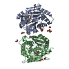 7twuC  4mikS S: Starting model for refinement C: citing same article ( |
|---|---|
| Similar structure data |
-Links
 PDBj
PDBj- Assembly
| Deposited unit | 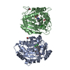
| ||||||||
|---|---|---|---|---|---|---|---|---|---|
| 1 |
| ||||||||
| Unit cell |
|
-Components
| #1: Protein | Mass: 32327.586 Da / Num. of mol.: 2 Source method: isolated from a genetically manipulated source Source: (gene. exp.) Homo sapiens (human) / Gene: PNMT, PENT / Production host:  References: UniProt: P11086, phenylethanolamine N-methyltransferase #2: Chemical |   Mass: 62.068 Da / Num. of mol.: 3 / Source method: obtained synthetically / Formula: C2H6O2 Mass: 62.068 Da / Num. of mol.: 3 / Source method: obtained synthetically / Formula: C2H6O2#3: Chemical |   Mass: 623.531 Da / Num. of mol.: 2 / Source method: obtained synthetically / Formula: C27H36Cl2N8O5 / Feature type: SUBJECT OF INVESTIGATION Mass: 623.531 Da / Num. of mol.: 2 / Source method: obtained synthetically / Formula: C27H36Cl2N8O5 / Feature type: SUBJECT OF INVESTIGATION#4: Water | ChemComp-HOH / |  Mass: 18.015 Da / Num. of mol.: 25 / Source method: isolated from a natural source / Formula: H2O Mass: 18.015 Da / Num. of mol.: 25 / Source method: isolated from a natural source / Formula: H2OHas ligand of interest | Y | Has protein modification | Y | |
|---|
-Experimental details
-Experiment
| Experiment | Method: X-RAY DIFFRACTION / Number of used crystals: 1 |
|---|
- Sample preparation
| Crystal | Density Matthews: 3.2 Å3/Da / Density % sol: 61.2 % |
|---|---|
| Crystal grow | Temperature: 295 K / Method: vapor diffusion, hanging drop / pH: 5.5 Details: 100 mM sodium cacodylate pH 5.5, 170 mM LiCl, 18 % PEG 6000 |
-Data collection
| Diffraction | Mean temperature: 100 K / Serial crystal experiment: N |
|---|---|
| Diffraction source | Source: SYNCHROTRON / Site: APS / Beamline: 31-ID / Wavelength: 0.9793 Å |
| Detector | Type: RAYONIX MX225HE / Detector: CCD / Date: Sep 16, 2021 |
| Radiation | Protocol: SINGLE WAVELENGTH / Monochromatic (M) / Laue (L): M / Scattering type: x-ray |
| Radiation wavelength | Wavelength: 0.9793 Å / Relative weight: 1 |
| Reflection | Resolution: 2.43→93.8 Å / Num. obs: 32221 / % possible obs: 100 % / Redundancy: 14.1 % / CC1/2: 1 / Rmerge(I) obs: 0.07 / Net I/σ(I): 24.2 |
| Reflection shell | Resolution: 2.43→2.52 Å / Redundancy: 13.9 % / Rmerge(I) obs: 1.62 / Mean I/σ(I) obs: 1.9 / Num. unique obs: 3321 / CC1/2: 0.81 / % possible all: 100 |
- Processing
| Software |
| ||||||||||||||||||||||||||||||||||||||||||||||||||||||||||||||||||||||||
|---|---|---|---|---|---|---|---|---|---|---|---|---|---|---|---|---|---|---|---|---|---|---|---|---|---|---|---|---|---|---|---|---|---|---|---|---|---|---|---|---|---|---|---|---|---|---|---|---|---|---|---|---|---|---|---|---|---|---|---|---|---|---|---|---|---|---|---|---|---|---|---|---|---|
| Refinement | Method to determine structure: MOLECULAR REPLACEMENT Starting model: 4MIK Resolution: 2.43→38.252 Å / SU ML: 0.34 / Cross valid method: THROUGHOUT / σ(F): 1.34 / Phase error: 32.07 / Stereochemistry target values: ML
| ||||||||||||||||||||||||||||||||||||||||||||||||||||||||||||||||||||||||
| Solvent computation | Shrinkage radii: 0.9 Å / VDW probe radii: 1.11 Å / Solvent model: FLAT BULK SOLVENT MODEL | ||||||||||||||||||||||||||||||||||||||||||||||||||||||||||||||||||||||||
| Displacement parameters | Biso max: 137.37 Å2 / Biso mean: 77.4427 Å2 / Biso min: 45.24 Å2 | ||||||||||||||||||||||||||||||||||||||||||||||||||||||||||||||||||||||||
| Refinement step | Cycle: final / Resolution: 2.43→38.252 Å
| ||||||||||||||||||||||||||||||||||||||||||||||||||||||||||||||||||||||||
| LS refinement shell | Refine-ID: X-RAY DIFFRACTION / Rfactor Rfree error: 0
|
