 Movie
Movie Controller

[English] 日本語
 Yorodumi
- PDB-7tvd: Crystal structure of the kinase domain of EGFR exon-19 (del-747-7... -
+ Open data

- Basic information
| Entry | Database: PDB / ID: 7tvd | ||||||
|---|---|---|---|---|---|---|---|
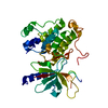
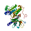
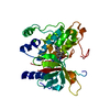
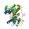
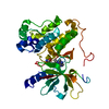
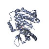
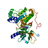
| Title | Crystal structure of the kinase domain of EGFR exon-19 (del-747-749) mutant | ||||||
 Components Components | Epidermal growth factor receptor | ||||||
 Keywords Keywords | TRANSFERASE / EGFR / exon-19 / kinase / ONCOPROTEIN | ||||||
| Function / homology |  Function and homology information Function and homology informationmultivesicular body, internal vesicle lumen / negative regulation of cardiocyte differentiation / Developmental Lineage of Mammary Gland Myoepithelial Cells / Shc-EGFR complex / positive regulation of protein kinase C signaling / Inhibition of Signaling by Overexpressed EGFR / epidermal growth factor receptor activity / EGFR interacts with phospholipase C-gamma / regulation of peptidyl-tyrosine phosphorylation / epidermal growth factor binding ...multivesicular body, internal vesicle lumen / negative regulation of cardiocyte differentiation / Developmental Lineage of Mammary Gland Myoepithelial Cells / Shc-EGFR complex / positive regulation of protein kinase C signaling / Inhibition of Signaling by Overexpressed EGFR / epidermal growth factor receptor activity / EGFR interacts with phospholipase C-gamma / regulation of peptidyl-tyrosine phosphorylation / epidermal growth factor binding / response to UV-A / ubiquitin-dependent endocytosis / PLCG1 events in ERBB2 signaling / ERBB2-EGFR signaling pathway / morphogenesis of an epithelial fold / PTK6 promotes HIF1A stabilization / Signaling by EGFR / ERBB2 Activates PTK6 Signaling / digestive tract morphogenesis / intracellular vesicle / eyelid development in camera-type eye / cerebral cortex cell migration / ERBB2 Regulates Cell Motility / protein insertion into membrane / negative regulation of epidermal growth factor receptor signaling pathway / protein tyrosine kinase activator activity / Respiratory syncytial virus (RSV) attachment and entry / Signaling by ERBB4 / PI3K events in ERBB2 signaling / positive regulation of phosphorylation / positive regulation of peptidyl-serine phosphorylation / hair follicle development / Estrogen-dependent nuclear events downstream of ESR-membrane signaling / MAP kinase kinase kinase activity / positive regulation of G1/S transition of mitotic cell cycle / GAB1 signalosome / embryonic placenta development / xenobiotic transport / salivary gland morphogenesis / positive regulation of epidermal growth factor receptor signaling pathway / sperm end piece / Signaling by ERBB2 / TFAP2 (AP-2) family regulates transcription of growth factors and their receptors / GRB2 events in EGFR signaling / transmembrane receptor protein tyrosine kinase activity / SHC1 events in EGFR signaling / EGFR Transactivation by Gastrin / GRB2 events in ERBB2 signaling / SHC1 events in ERBB2 signaling / sperm principal piece / ossification / basal plasma membrane / cellular response to epidermal growth factor stimulus / epithelial cell proliferation / positive regulation of DNA replication / positive regulation of DNA repair / positive regulation of epithelial cell proliferation / Signal transduction by L1 / positive regulation of protein localization to plasma membrane / cellular response to amino acid stimulus / phosphatidylinositol 3-kinase/protein kinase B signal transduction / NOTCH3 Activation and Transmission of Signal to the Nucleus / cellular response to estradiol stimulus / clathrin-coated endocytic vesicle membrane / EGFR downregulation / Signaling by ERBB2 TMD/JMD mutants / cell-cell adhesion / Constitutive Signaling by EGFRvIII / receptor protein-tyrosine kinase / Signaling by ERBB2 ECD mutants / negative regulation of protein catabolic process / Signaling by ERBB2 KD Mutants / positive regulation of miRNA transcription / epidermal growth factor receptor signaling pathway / kinase binding / ruffle membrane / Downregulation of ERBB2 signaling / positive regulation of fibroblast proliferation / cell morphogenesis / positive regulation of protein phosphorylation / neuron differentiation / Constitutive Signaling by Aberrant PI3K in Cancer / HCMV Early Events / actin filament binding / cell junction / transmembrane signaling receptor activity / positive regulation of canonical Wnt signaling pathway / sperm midpiece / PIP3 activates AKT signaling / Cargo recognition for clathrin-mediated endocytosis / Constitutive Signaling by Ligand-Responsive EGFR Cancer Variants / Clathrin-mediated endocytosis / ATPase binding / PI5P, PP2A and IER3 Regulate PI3K/AKT Signaling / virus receptor activity / RAF/MAP kinase cascade / positive regulation of cell growth / protein tyrosine kinase activity / double-stranded DNA binding / early endosome membrane Similarity search - Function | ||||||
| Biological species |  Homo sapiens (human) Homo sapiens (human) | ||||||
| Method |  X-RAY DIFFRACTION / SYNCHROTRON / MOLECULAR REPLACEMENT / Resolution: 2.96 Å X-RAY DIFFRACTION / SYNCHROTRON / MOLECULAR REPLACEMENT / Resolution: 2.96 Å | ||||||
 Authors Authors | Ashtekar, K.D. / Stayrook, S.E. / Lemmon, M.A. | ||||||
| Funding support |  United States, 1items United States, 1items
| ||||||
 Citation Citation | Journal: Nat Commun / Year: 2022 Title: Biochemical and structural basis for differential inhibitor sensitivity of EGFR with distinct exon 19 mutations. Authors: van Alderwerelt van Rosenburgh, I.K. / Lu, D.M. / Grant, M.J. / Stayrook, S.E. / Phadke, M. / Walther, Z. / Goldberg, S.B. / Politi, K. / Lemmon, M.A. / Ashtekar, K.D. / Tsutsui, Y. | ||||||
| History |
|
- Structure visualization
| Structure viewer | Molecule: MolmilJmol/JSmol |
|---|
- Downloads & links
-Download
| PDBx/mmCIF format | 7tvd.cif.gz | 157.6 KB | Display | PDBx/mmCIF format |
|---|---|---|---|---|
| PDB format | pdb7tvd.ent.gz | 104.1 KB | Display | PDB format |
| PDBx/mmJSON format | 7tvd.json.gz | Tree view | PDBx/mmJSON format | |
| Others |  Other downloads Other downloads |
-Validation report
| Arichive directory | https://data.pdbj.org/pub/pdb/validation_reports/tv/7tvdftp://data.pdbj.org/pub/pdb/validation_reports/tv/7tvd | HTTPS FTP |
|---|
-Related structure data
| Related structure data | 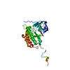 1m17S S: Starting model for refinement |
|---|---|
| Similar structure data |


-Links
 PDBj
PDBj


- Assembly
| Deposited unit | 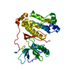
| ||||||||||||
|---|---|---|---|---|---|---|---|---|---|---|---|---|---|
| 1 |
| ||||||||||||
| Unit cell |
|
-Components
| #1: Protein | Mass: 38237.191 Da / Num. of mol.: 1 Source method: isolated from a genetically manipulated source Source: (gene. exp.) Homo sapiens (human) / Gene: EGFR, ERBB, ERBB1, HER1 / Production host:   Spodoptera frugiperda (fall armyworm) Spodoptera frugiperda (fall armyworm)References: UniProt: P00533, receptor protein-tyrosine kinase |
|---|
-Experimental details
-Experiment
| Experiment | Method: X-RAY DIFFRACTION / Number of used crystals: 1 |
|---|
- Sample preparation
| Crystal | Density Matthews: 3.64 Å3/Da / Density % sol: 66.16 % |
|---|---|
| Crystal grow | Temperature: 289.15 K / Method: vapor diffusion, hanging drop / pH: 7.5 Details: 28% PEG 400, 0.1 M Na Hepes pH 7.5, 0.2 M Calcium Chloride |
-Data collection
| Diffraction | Mean temperature: 100 K / Serial crystal experiment: N | |||||||||||||||||||||||||||
|---|---|---|---|---|---|---|---|---|---|---|---|---|---|---|---|---|---|---|---|---|---|---|---|---|---|---|---|---|
| Diffraction source | Source: SYNCHROTRON / Site: APS / Beamline: 24-ID-C / Wavelength: 0.97918 Å | |||||||||||||||||||||||||||
| Detector | Type: DECTRIS EIGER X 16M / Detector: PIXEL / Date: Mar 19, 2019 / Details: undulator | |||||||||||||||||||||||||||
| Radiation | Protocol: SINGLE WAVELENGTH / Monochromatic (M) / Laue (L): M / Scattering type: x-ray | |||||||||||||||||||||||||||
| Radiation wavelength | Wavelength: 0.97918 Å / Relative weight: 1 | |||||||||||||||||||||||||||
| Reflection | Resolution: 2.96→105.66 Å / Num. obs: 11749 / % possible obs: 100 % / Redundancy: 36.1 % / CC1/2: 0.999 / Rmerge(I) obs: 0.259 / Rpim(I) all: 0.044 / Rrim(I) all: 0.263 / Net I/σ(I): 15.4 / Num. measured all: 423785 | |||||||||||||||||||||||||||
| Reflection shell | Diffraction-ID: 1 / % possible all: 100
|
- Processing
| Software |
| ||||||||||||||||||||||||||||||||||||||||
|---|---|---|---|---|---|---|---|---|---|---|---|---|---|---|---|---|---|---|---|---|---|---|---|---|---|---|---|---|---|---|---|---|---|---|---|---|---|---|---|---|---|
| Refinement | Method to determine structure: MOLECULAR REPLACEMENT Starting model: 1M17 Resolution: 2.96→74.71 Å / SU ML: 0.4085 / Cross valid method: FREE R-VALUE / σ(F): 1.34 / Phase error: 35.9126 Stereochemistry target values: GeoStd + Monomer Library + CDL v1.2
| ||||||||||||||||||||||||||||||||||||||||
| Solvent computation | Shrinkage radii: 0.9 Å / VDW probe radii: 1.11 Å / Solvent model: FLAT BULK SOLVENT MODEL | ||||||||||||||||||||||||||||||||||||||||
| Displacement parameters | Biso mean: 104.92 Å2 | ||||||||||||||||||||||||||||||||||||||||
| Refinement step | Cycle: LAST / Resolution: 2.96→74.71 Å
| ||||||||||||||||||||||||||||||||||||||||
| Refine LS restraints |
| ||||||||||||||||||||||||||||||||||||||||
| LS refinement shell |
| ||||||||||||||||||||||||||||||||||||||||
| Refinement TLS params. | Method: refined / Origin x: -60.3983808351 Å / Origin y: 7.60852047851 Å / Origin z: 25.513823797 Å
| ||||||||||||||||||||||||||||||||||||||||
| Refinement TLS group | Selection details: all |
