Entry Database : PDB / ID : 7tvaTitle Stat5a Core in complex with AK2292 Signal transducer and activator of transcription 5A Keywords / / Function / homology Function Domain/homology Component
/ / / / / / / / / / / / / / / / / / / / / / / / / / / / / / / / / / / / / / / / / / / / / / / / / / / / / / / / / / / / / / / / / / / / / / / / / / / / / / / / / / / / / / / / / / / / / / / / / / / / / / / / / / / / / / / / / / / / / / / / / / / / / / / / / / / / / / / / / / / / / / Biological species Homo sapiens (human)Method / / / / Resolution : 2.835 Å Authors Meagher, J.L. / Stuckey, J.A. Funding support Organization Grant number Country National Institutes of Health/National Cancer Institute (NIH/NCI) 1-R01-CA244509
Journal : Nat.Chem.Biol. / Year : 2023Title : A selective small-molecule STAT5 PROTAC degrader capable of achieving tumor regression in vivo.Authors: Kaneshige, A. / Bai, L. / Wang, M. / McEachern, D. / Meagher, J.L. / Xu, R. / Wang, Y. / Jiang, W. / Metwally, H. / Kirchhoff, P.D. / Zhao, L. / Jiang, H. / Wang, M. / Wen, B. / Sun, D. / ... Authors : Kaneshige, A. / Bai, L. / Wang, M. / McEachern, D. / Meagher, J.L. / Xu, R. / Wang, Y. / Jiang, W. / Metwally, H. / Kirchhoff, P.D. / Zhao, L. / Jiang, H. / Wang, M. / Wen, B. / Sun, D. / Stuckey, J.A. / Wang, S. History Deposition Feb 4, 2022 Deposition site / Processing site Revision 1.0 Feb 15, 2023 Provider / Type Revision 1.1 Jun 7, 2023 Group / Category Item / _citation.page_first / _citation.page_lastRevision 1.2 Apr 3, 2024 Group / Refinement descriptionCategory / chem_comp_bond / pdbx_initial_refinement_model
Show all Show less
 Movie
Movie Controller


 Open data
 Basic information
 Components
 Keywords
 Function and homology information
 Homo sapiens (human)
 X-RAY DIFFRACTION /
  Authors
 United States, 1items
  Citation
 Structure visualization
 Downloads & links
 Other downloads
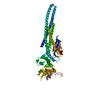
 PDBj
PDBj


 Assembly
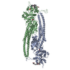


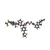
 Mass: 1070.127 Da / Num. of mol.: 2 / Source method: obtained synthetically / Formula: C52H54F2N7O10PS2 / Feature type: SUBJECT OF INVESTIGATION
 Mass: 106.120 Da / Num. of mol.: 2 / Source method: obtained synthetically / Formula: C4H10O3
 Mass: 102.046 Da / Num. of mol.: 1 / Source method: obtained synthetically / Formula: C3H2O4
 Mass: 18.015 Da / Num. of mol.: 232 / Source method: isolated from a natural source / Formula: H2O
 Sample preparation
 Processing