- PDB-7tul: Woodchuck hepatitis small surface protein without cytosolic and a... -
+
Open data
ID or keywords:
Loading...
-
Basic information
Entry
Database: PDB / ID: 7tul
Title
Woodchuck hepatitis small surface protein without cytosolic and antigenic loops
Components
Large envelope protein
Keywords
VIRAL PROTEIN / WHV / SVP / WHsAg / envelope protein
Function / homology
Large envelope protein S / Major surface antigen from hepadnavirus / caveolin-mediated endocytosis of virus by host cell / fusion of virus membrane with host endosome membrane / viral envelope / virion attachment to host cell / virion membrane / membrane / Large envelope protein
Function and homology information
Biological species
Woodchuck hepatitis virus
Method
ELECTRON MICROSCOPY / single particle reconstruction / cryo EM / Resolution: 6.5 Å
National Institutes of Health/National Institute Of Allergy and Infectious Diseases (NIH/NIAID)
United States
Citation
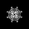
Journal: Sci Adv / Year: 2022 Title: Cryo-EM structures of human hepatitis B and woodchuck hepatitis virus small spherical subviral particles. Authors: Haitao Liu / Xupeng Hong / Ji Xi / Stephan Menne / Jianming Hu / Joseph Che-Yen Wang / Abstract: The loss of detectable hepatitis B surface antigen (HBsAg) is considered a functional cure in chronic hepatitis B. Naturally, HBsAg can be incorporated into the virion envelope or assembled into ...The loss of detectable hepatitis B surface antigen (HBsAg) is considered a functional cure in chronic hepatitis B. Naturally, HBsAg can be incorporated into the virion envelope or assembled into subviral particles (SVPs) with lipid from host cells. Until now, there has been no detailed structure of HBsAg, and the published SVP structures are controversial. Here, we report the first subnanometer-resolution structures of spherical SVP from hepatitis B virus (HBV) and the related woodchuck hepatitis virus (WHV) determined by cryo-electron microscopy in combination with AlphaFold2 prediction. Both structures showed unique rhombicuboctahedral symmetry with 24 protruding spikes comprising dimer of small HBsAg with four helical domains. The lipid moiety in the SVP is organized in a noncanonical lipid patch instead of a lipid bilayer, which can accommodate the exposed hydrophobic surface and modulate particle stability. Together, these findings advance our knowledge of viral membrane organization and the structures of HBV and WHV spherical SVPs.
History
Deposition
Feb 2, 2022
Deposition site: RCSB / Processing site: RCSB
Revision 1.0
Aug 24, 2022
Provider: repository / Type: Initial release
Revision 1.1
Feb 21, 2024
Group: Data collection / Category: chem_comp_atom / chem_comp_bond
Electron dose: 30 e/Å2 / Film or detector model: GATAN K3 BIOQUANTUM (6k x 4k)
-
Processing
CTF correction
Type: PHASE FLIPPING AND AMPLITUDE CORRECTION
3D reconstruction
Resolution: 6.5 Å / Resolution method: FSC 0.143 CUT-OFF / Num. of particles: 34520 / Symmetry type: POINT
Atomic model building
Details: The authors state that the model was built using 6.5 A cryo-EM map and Alphafold2, so the clashes cannot be totally resolved due to the lack of density from cryo-EM maps.
+
About Yorodumi
-
News
-
Feb 9, 2022. New format data for meta-information of EMDB entries
New format data for meta-information of EMDB entries
Version 3 of the EMDB header file is now the official format.
The previous official version 1.9 will be removed from the archive.
In the structure databanks used in Yorodumi, some data are registered as the other names, "COVID-19 virus" and "2019-nCoV". Here are the details of the virus and the list of structure data.
Jan 31, 2019. EMDB accession codes are about to change! (news from PDBe EMDB page)
EMDB accession codes are about to change! (news from PDBe EMDB page)
The allocation of 4 digits for EMDB accession codes will soon come to an end. Whilst these codes will remain in use, new EMDB accession codes will include an additional digit and will expand incrementally as the available range of codes is exhausted. The current 4-digit format prefixed with “EMD-” (i.e. EMD-XXXX) will advance to a 5-digit format (i.e. EMD-XXXXX), and so on. It is currently estimated that the 4-digit codes will be depleted around Spring 2019, at which point the 5-digit format will come into force.
The EM Navigator/Yorodumi systems omit the EMD- prefix.
Related info.:Q: What is EMD? / ID/Accession-code notation in Yorodumi/EM Navigator
Yorodumi is a browser for structure data from EMDB, PDB, SASBDB, etc.
This page is also the successor to EM Navigator detail page, and also detail information page/front-end page for Omokage search.
The word "yorodu" (or yorozu) is an old Japanese word meaning "ten thousand". "mi" (miru) is to see.
Related info.:EMDB / PDB / SASBDB / Comparison of 3 databanks / Yorodumi Search / Aug 31, 2016. New EM Navigator & Yorodumi / Yorodumi Papers / Jmol/JSmol / Function and homology information / Changes in new EM Navigator and Yorodumi
 Movie
Movie Controller

 Yorodumi
 Open data
 Basic information
 Components
 Keywords
 Function and homology information
 Woodchuck hepatitis virus
 Authors
 United States, 1items
  Citation
 Structure visualization
 Downloads & links
 Other downloads
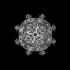


 PDBj
PDBj Assembly
 Sample preparation
 Electron microscopy imaging
 FIELD EMISSION GUN / Accelerating voltage: 300 kV / Illumination mode: FLOOD BEAM
 Processing