 Movie
Movie Controller

[English] 日本語
 Yorodumi
- PDB-7tuk: Small hepatitis B virus surface protein without cytosolic and ant... -
+ Open data

- Basic information
| Entry | Database: PDB / ID: 7tuk | ||||||
|---|---|---|---|---|---|---|---|
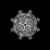
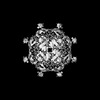
| Title | Small hepatitis B virus surface protein without cytosolic and antigenic loops | ||||||
 Components Components | SAg protein | ||||||
 Keywords Keywords | VIRAL PROTEIN / HBV / SVP / HBsAg / Envelope protein | ||||||
| Function / homology | Large envelope protein S / Major surface antigen from hepadnavirus / caveolin-mediated endocytosis of virus by host cell / fusion of virus membrane with host endosome membrane / viral envelope / membrane / SAg protein Function and homology information Function and homology information | ||||||
| Biological species |  HBV genotype E (virus) HBV genotype E (virus) | ||||||
| Method | ELECTRON MICROSCOPY / single particle reconstruction / cryo EM / Resolution: 6.3 Å | ||||||
 Authors Authors | Liu, H. / Wang, J.C.Y. | ||||||
| Funding support |  United States, 1items United States, 1items
| ||||||
 Citation Citation | Journal: Sci Adv / Year: 2022 Title: Cryo-EM structures of human hepatitis B and woodchuck hepatitis virus small spherical subviral particles. Authors: Haitao Liu / Xupeng Hong / Ji Xi / Stephan Menne / Jianming Hu / Joseph Che-Yen Wang / Abstract: The loss of detectable hepatitis B surface antigen (HBsAg) is considered a functional cure in chronic hepatitis B. Naturally, HBsAg can be incorporated into the virion envelope or assembled into ...The loss of detectable hepatitis B surface antigen (HBsAg) is considered a functional cure in chronic hepatitis B. Naturally, HBsAg can be incorporated into the virion envelope or assembled into subviral particles (SVPs) with lipid from host cells. Until now, there has been no detailed structure of HBsAg, and the published SVP structures are controversial. Here, we report the first subnanometer-resolution structures of spherical SVP from hepatitis B virus (HBV) and the related woodchuck hepatitis virus (WHV) determined by cryo-electron microscopy in combination with AlphaFold2 prediction. Both structures showed unique rhombicuboctahedral symmetry with 24 protruding spikes comprising dimer of small HBsAg with four helical domains. The lipid moiety in the SVP is organized in a noncanonical lipid patch instead of a lipid bilayer, which can accommodate the exposed hydrophobic surface and modulate particle stability. Together, these findings advance our knowledge of viral membrane organization and the structures of HBV and WHV spherical SVPs. | ||||||
| History |
|
- Structure visualization
| Structure viewer | Molecule: MolmilJmol/JSmol |
|---|
- Downloads & links
-Download
| PDBx/mmCIF format | 7tuk.cif.gz | 110.9 KB | Display | PDBx/mmCIF format |
|---|---|---|---|---|
| PDB format | pdb7tuk.ent.gz | 86.9 KB | Display | PDB format |
| PDBx/mmJSON format | 7tuk.json.gz | Tree view | PDBx/mmJSON format | |
| Others |  Other downloads Other downloads |
-Validation report
| Arichive directory | https://data.pdbj.org/pub/pdb/validation_reports/tu/7tukftp://data.pdbj.org/pub/pdb/validation_reports/tu/7tuk | HTTPS FTP |
|---|
-Related structure data
| Related structure data |  26117MC  7tulC M: map data used to model this data C: citing same article ( |
|---|---|
| Similar structure data |
-Links
 PDBj
PDBj- Assembly
| Deposited unit | 
|
|---|---|
| 1 |
|
-Components
| #1: Protein | Mass: 25303.992 Da / Num. of mol.: 2 Source method: isolated from a genetically manipulated source Source: (gene. exp.) HBV genotype E (virus) / Gene: S / Production host:  Homo sapiens (human) / References: UniProt: D6CIG5 Homo sapiens (human) / References: UniProt: D6CIG5 |
|---|
-Experimental details
-Experiment
| Experiment | Method: ELECTRON MICROSCOPY |
|---|---|
| EM experiment | Aggregation state: PARTICLE / 3D reconstruction method: single particle reconstruction |
- Sample preparation
| Component | Name: HBV genotype E / Type: VIRUS / Entity ID: all / Source: NATURAL |
|---|---|
| Source (natural) | Organism: HBV genotype E (virus) |
| Details of virus | Empty: YES / Enveloped: YES / Isolate: STRAIN / Type: VIRION |
| Buffer solution | pH: 8 |
| Specimen | Embedding applied: NO / Shadowing applied: NO / Staining applied: NO / Vitrification applied: YES |
| Specimen support | Grid material: COPPER / Grid mesh size: 300 divisions/in. / Grid type: Quantifoil R2/2 |
| Vitrification | Instrument: FEI VITROBOT MARK IV / Cryogen name: ETHANE / Humidity: 100 % |
- Electron microscopy imaging
| Experimental equipment |  Model: Titan Krios / Image courtesy: FEI Company |
|---|---|
| Microscopy | Model: TFS KRIOS |
| Electron gun | Electron source:  FIELD EMISSION GUN / Accelerating voltage: 300 kV / Illumination mode: FLOOD BEAM FIELD EMISSION GUN / Accelerating voltage: 300 kV / Illumination mode: FLOOD BEAM |
| Electron lens | Mode: BRIGHT FIELD / Nominal defocus max: 3200 nm / Nominal defocus min: 800 nm / Cs: 2.7 mm / C2 aperture diameter: 50 µm |
| Image recording | Average exposure time: 3 sec. / Electron dose: 30 e/Å2 / Film or detector model: GATAN K3 (6k x 4k) / Num. of grids imaged: 1 |
- Processing
| CTF correction | Type: PHASE FLIPPING AND AMPLITUDE CORRECTION |
|---|---|
| 3D reconstruction | Resolution: 6.3 Å / Resolution method: FSC 0.143 CUT-OFF / Num. of particles: 36166 / Symmetry type: POINT |
| Atomic model building | Details: The authors state that the model was built using 6.5 A cryo-EM map and Alphafold2, so the clashes cannot be totally resolved due to the lack of density from cryo-EM maps. |
