 Movie
Movie Controller

[English] 日本語
 Yorodumi
- PDB-7tmf: Crystal Structure of NAD(P)H nitroreductase from Klebsiella pneum... -
+ Open data

- Basic information
| Entry | Database: PDB / ID: 7tmf | ||||||
|---|---|---|---|---|---|---|---|
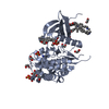
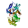
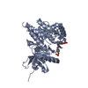
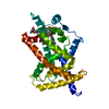
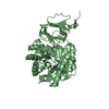
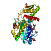
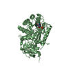
| Title | Crystal Structure of NAD(P)H nitroreductase from Klebsiella pneumoniae (short b-axis) | ||||||
 Components Components | NAD(P)H nitroreductase | ||||||
 Keywords Keywords | OXIDOREDUCTASE / nitroreductase / SSGCID / STRUCTURAL GENOMICS / SEATTLE STRUCTURAL GENOMICS CENTER FOR INFECTIOUS DISEASE | ||||||
| Function / homology |  Function and homology information Function and homology information | ||||||
| Biological species |  Klebsiella pneumoniae subsp. pneumoniae HS11286 (bacteria) Klebsiella pneumoniae subsp. pneumoniae HS11286 (bacteria) | ||||||
| Method |  X-RAY DIFFRACTION / SYNCHROTRON / MOLECULAR REPLACEMENT / molecular replacement / Resolution: 1.97 Å X-RAY DIFFRACTION / SYNCHROTRON / MOLECULAR REPLACEMENT / molecular replacement / Resolution: 1.97 Å | ||||||
 Authors Authors | Seattle Structural Genomics Center for Infectious Disease (SSGCID) | ||||||
| Funding support |  United States, 1items United States, 1items
| ||||||
 Citation Citation | Journal: Acta Crystallogr.,Sect.F / Year: 2024 Title: Crystal structures of NAD(P)H nitroreductases from Klebsiella pneumoniae Authors: Kancherla, A.D. / Liu, L. / Tillery, L. / Shek, R. / Craig, J.K. / Machen, A.J. / Seibold, S. / Battaile, K.P. / Fradi, S. / Barrett, L.K. / Subramanian, S. / Myler, P. / Van Voorhis, W.C. / Lovell, S. | ||||||
| History |
|
- Structure visualization
| Structure viewer | Molecule: MolmilJmol/JSmol |
|---|
- Downloads & links
-Download
| PDBx/mmCIF format | 7tmf.cif.gz | 90.6 KB | Display | PDBx/mmCIF format |
|---|---|---|---|---|
| PDB format | pdb7tmf.ent.gz | 66.3 KB | Display | PDB format |
| PDBx/mmJSON format | 7tmf.json.gz | Tree view | PDBx/mmJSON format | |
| Others |  Other downloads Other downloads |
-Validation report
| Summary document | 7tmf_validation.pdf.gz | 1 MB | Display | wwPDB validaton report |
|---|---|---|---|---|
| Full document | 7tmf_full_validation.pdf.gz | 1.1 MB | Display | |
| Data in XML | 7tmf_validation.xml.gz | 17.1 KB | Display | |
| Data in CIF | 7tmf_validation.cif.gz | 23.4 KB | Display | |
| Arichive directory | https://data.pdbj.org/pub/pdb/validation_reports/tm/7tmfftp://data.pdbj.org/pub/pdb/validation_reports/tm/7tmf | HTTPS FTP |
-Related structure data
| Related structure data | 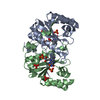 7tmgC  3bm1S S: Starting model for refinement C: citing same article ( |
|---|---|
| Similar structure data |

-Links
 PDBj
PDBj- Assembly
| Deposited unit | 
| ||||||||
|---|---|---|---|---|---|---|---|---|---|
| 1 |
| ||||||||
| Unit cell |
|
-Components
-Protein , 1 types, 2 molecules AB
| #1: Protein | Mass: 21245.316 Da / Num. of mol.: 2 / Fragment: KlpnC.17456.a.B1 Source method: isolated from a genetically manipulated source Source: (gene. exp.) Klebsiella pneumoniae subsp. pneumoniae HS11286 (bacteria)Gene: ydjA, E3U37_20475, E3U38_02825, E9161_16570, NCTC9504_03979 Plasmid: KlpnC.17456.a.B1 / Production host: |
|---|
-Non-polymers , 6 types, 156 molecules 


| #2: Chemical |  Mass: 456.344 Da / Num. of mol.: 2 / Source method: obtained synthetically / Formula: C17H21N4O9P / Feature type: SUBJECT OF INVESTIGATION Mass: 456.344 Da / Num. of mol.: 2 / Source method: obtained synthetically / Formula: C17H21N4O9P / Feature type: SUBJECT OF INVESTIGATION#3: Chemical | ChemComp-BTB / |  Mass: 209.240 Da / Num. of mol.: 1 / Source method: obtained synthetically / Formula: C8H19NO5 / Comment: pH buffer*YM Mass: 209.240 Da / Num. of mol.: 1 / Source method: obtained synthetically / Formula: C8H19NO5 / Comment: pH buffer*YM#4: Chemical |  Mass: 92.094 Da / Num. of mol.: 3 / Source method: obtained synthetically / Formula: C3H8O3 Mass: 92.094 Da / Num. of mol.: 3 / Source method: obtained synthetically / Formula: C3H8O3#5: Chemical | ChemComp-PO4 /  Mass: 94.971 Da / Num. of mol.: 9 / Source method: obtained synthetically / Formula: PO4 Mass: 94.971 Da / Num. of mol.: 9 / Source method: obtained synthetically / Formula: PO4#6: Chemical | ChemComp-1PE / |  Mass: 238.278 Da / Num. of mol.: 1 / Source method: obtained synthetically / Formula: C10H22O6 / Comment: precipitant*YM Mass: 238.278 Da / Num. of mol.: 1 / Source method: obtained synthetically / Formula: C10H22O6 / Comment: precipitant*YM#7: Water | ChemComp-HOH / | Mass: 18.015 Da / Num. of mol.: 140 / Source method: isolated from a natural source / Formula: H2O |
|---|
-Details
| Has ligand of interest | Y |
|---|
-Experimental details
-Experiment
| Experiment | Method: X-RAY DIFFRACTION / Number of used crystals: 1 |
|---|
- Sample preparation
| Crystal | Density Matthews: 2.37 Å3/Da / Density % sol: 48.1 % / Mosaicity: 0.06 ° |
|---|---|
| Crystal grow | Temperature: 291 K / Method: vapor diffusion, sitting drop / pH: 6.5 Details: Berkeley C1: 25 %w/v PEG 3350, 0.2 M (NH4)2SO4, 0.1 M BIS-TRIS, KlpnC.17456.a.B1.PW39057 at 21 mg/mL, Tray: plate 12177 well C1 drop 1, Puck: PSL0413, Cryo: 80% crystallant and 20% PEG 200, |
-Data collection
| Diffraction | Mean temperature: 100 K / Serial crystal experiment: N | ||||||||||||||||||||||||||||||
|---|---|---|---|---|---|---|---|---|---|---|---|---|---|---|---|---|---|---|---|---|---|---|---|---|---|---|---|---|---|---|---|
| Diffraction source | Source: SYNCHROTRON / Site: NSLS-II / Beamline: 19-ID / Wavelength: 0.9795 Å | ||||||||||||||||||||||||||||||
| Detector | Type: DECTRIS PILATUS 6M / Detector: PIXEL / Date: Oct 19, 2021 | ||||||||||||||||||||||||||||||
| Radiation | Protocol: SINGLE WAVELENGTH / Monochromatic (M) / Laue (L): M / Scattering type: x-ray | ||||||||||||||||||||||||||||||
| Radiation wavelength | Wavelength: 0.9795 Å / Relative weight: 1 | ||||||||||||||||||||||||||||||
| Reflection | Resolution: 1.97→92.22 Å / Num. obs: 29253 / % possible obs: 100 % / Redundancy: 6.4 % / Biso Wilson estimate: 29.83 Å2 / CC1/2: 0.998 / Rmerge(I) obs: 0.099 / Rpim(I) all: 0.042 / Rrim(I) all: 0.107 / Net I/σ(I): 12.4 / Num. measured all: 188186 / Scaling rejects: 59 | ||||||||||||||||||||||||||||||
| Reflection shell | Diffraction-ID: 1
|
-Phasing
| Phasing | Method: molecular replacement |
|---|
- Processing
| Software |
| |||||||||||||||||||||||||||||||||||||||||||||||||||||||||||||||||||||||||||||
|---|---|---|---|---|---|---|---|---|---|---|---|---|---|---|---|---|---|---|---|---|---|---|---|---|---|---|---|---|---|---|---|---|---|---|---|---|---|---|---|---|---|---|---|---|---|---|---|---|---|---|---|---|---|---|---|---|---|---|---|---|---|---|---|---|---|---|---|---|---|---|---|---|---|---|---|---|---|---|
| Refinement | Method to determine structure: MOLECULAR REPLACEMENT Starting model: 3BM1 Resolution: 1.97→55.17 Å / SU ML: 0.25 / Cross valid method: THROUGHOUT / σ(F): 1.34 / Phase error: 24.31 / Stereochemistry target values: ML
| |||||||||||||||||||||||||||||||||||||||||||||||||||||||||||||||||||||||||||||
| Solvent computation | Shrinkage radii: 0.9 Å / VDW probe radii: 1.11 Å / Solvent model: FLAT BULK SOLVENT MODEL | |||||||||||||||||||||||||||||||||||||||||||||||||||||||||||||||||||||||||||||
| Displacement parameters | Biso max: 85.55 Å2 / Biso mean: 31.4906 Å2 / Biso min: 17.91 Å2 | |||||||||||||||||||||||||||||||||||||||||||||||||||||||||||||||||||||||||||||
| Refinement step | Cycle: final / Resolution: 1.97→55.17 Å
| |||||||||||||||||||||||||||||||||||||||||||||||||||||||||||||||||||||||||||||
| LS refinement shell | Refine-ID: X-RAY DIFFRACTION / Rfactor Rfree error: 0 / Total num. of bins used: 10
|
