 Movie
Movie Controller

[English] 日本語
 Yorodumi
- PDB-7tl8: 1.95A resolution structure of independent phosphoglycerate mutase... -
+ Open data

- Basic information
| Entry | Database: PDB / ID: 7tl8 | ||||||
|---|---|---|---|---|---|---|---|
| Title | 1.95A resolution structure of independent phosphoglycerate mutase from S. aureus in complex with a macrocyclic peptide inhibitor (Sa-D3) | ||||||
 Components Components |
| ||||||
 Keywords Keywords | ISOMERASE/Inhibitor / phosphoglycerate mutase / ipglycermide / macrocyclic peptide inhibitors / metal ion binding / ISOMERASE / ISOMERASE-Inhibitor complex | ||||||
| Function / homology |  Function and homology information Function and homology information: / phosphoglycerate mutase (2,3-diphosphoglycerate-independent) / glucose catabolic process / glycolytic process / manganese ion binding / cytoplasm Similarity search - Function | ||||||
| Biological species |   Staphylococcus aureus (bacteria) Staphylococcus aureus (bacteria)synthetic construct (others) | ||||||
| Method |  X-RAY DIFFRACTION / SYNCHROTRON / MOLECULAR REPLACEMENT / molecular replacement / Resolution: 1.95 Å X-RAY DIFFRACTION / SYNCHROTRON / MOLECULAR REPLACEMENT / molecular replacement / Resolution: 1.95 Å | ||||||
 Authors Authors | Liu, L. / Lovell, S. / Battaile, K.P. / Dranchak, P. / Queme, B. / Aitha, M. / van Neer, R.H.P. / Kimura, H. / Katho, T. / Suga, H. / Inglese, J. | ||||||
| Funding support | 1items
| ||||||
 Citation Citation | Journal: Acs Chem.Biol. / Year: 2022 Title: Serum-Stable and Selective Backbone-N-Methylated Cyclic Peptides That Inhibit Prokaryotic Glycolytic Mutases. Authors: van Neer, R.H.P. / Dranchak, P.K. / Liu, L. / Aitha, M. / Queme, B. / Kimura, H. / Katoh, T. / Battaile, K.P. / Lovell, S. / Inglese, J. / Suga, H. | ||||||
| History |
|
- Structure visualization
| Structure viewer | Molecule: MolmilJmol/JSmol |
|---|
- Downloads & links
-Download
| PDBx/mmCIF format | 7tl8.cif.gz | 119.1 KB | Display | PDBx/mmCIF format |
|---|---|---|---|---|
| PDB format | pdb7tl8.ent.gz | 87.3 KB | Display | PDB format |
| PDBx/mmJSON format | 7tl8.json.gz | Tree view | PDBx/mmJSON format | |
| Others |  Other downloads Other downloads |
-Validation report
| Summary document | 7tl8_validation.pdf.gz | 434.5 KB | Display | wwPDB validaton report |
|---|---|---|---|---|
| Full document | 7tl8_full_validation.pdf.gz | 436.1 KB | Display | |
| Data in XML | 7tl8_validation.xml.gz | 21.1 KB | Display | |
| Data in CIF | 7tl8_validation.cif.gz | 30.5 KB | Display | |
| Arichive directory | https://data.pdbj.org/pub/pdb/validation_reports/tl/7tl8ftp://data.pdbj.org/pub/pdb/validation_reports/tl/7tl8 | HTTPS FTP |
-Related structure data
| Related structure data |  7tl7C 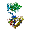 4my4S S: Starting model for refinement C: citing same article ( |
|---|---|
| Similar structure data |
-Links
 PDBj
PDBj
- Assembly
| Deposited unit | 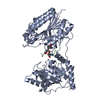
| ||||||||
|---|---|---|---|---|---|---|---|---|---|
| 1 |
| ||||||||
| Unit cell |
|
-Components
| #1: Protein | Mass: 57316.688 Da / Num. of mol.: 1 Source method: isolated from a genetically manipulated source Source: (gene. exp.) Staphylococcus aureus (bacteria) / Gene: pgm_1, gpmI, pgm_2 / Plasmid: pET21a(+) / Production host: References: UniProt: W8U5L7, phosphoglycerate mutase (2,3-diphosphoglycerate-independent) | ||||
|---|---|---|---|---|---|
| #2: Protein/peptide | Type: Cyclic peptide / Class: Inhibitor / Mass: 1809.991 Da / Num. of mol.: 1 / Source method: obtained synthetically / Source: (synth.) synthetic construct (others) / References: BIRD: PRD_002492 | ||||
| #3: Chemical |   Mass: 54.938 Da / Num. of mol.: 2 / Source method: obtained synthetically / Formula: Mn Mass: 54.938 Da / Num. of mol.: 2 / Source method: obtained synthetically / Formula: Mn#4: Water | ChemComp-HOH / |  Mass: 18.015 Da / Num. of mol.: 231 / Source method: isolated from a natural source / Formula: H2O Mass: 18.015 Da / Num. of mol.: 231 / Source method: isolated from a natural source / Formula: H2OHas ligand of interest | Y | |
-Experimental details
-Experiment
| Experiment | Method: X-RAY DIFFRACTION / Number of used crystals: 1 |
|---|
- Sample preparation
| Crystal | Density Matthews: 2.17 Å3/Da / Density % sol: 43.3 % / Mosaicity: 0.19 ° |
|---|---|
| Crystal grow | Temperature: 291 K / Method: vapor diffusion, sitting drop / pH: 6.5 Details: 25% (w/v) PEG 3350, 100 mM Bis-Tris, 200 mM ammonium acetate |
-Data collection
| Diffraction | Mean temperature: 100 K / Serial crystal experiment: N | ||||||||||||||||||||||||
|---|---|---|---|---|---|---|---|---|---|---|---|---|---|---|---|---|---|---|---|---|---|---|---|---|---|
| Diffraction source | Source: SYNCHROTRON / Site: NSLS-II  / Beamline: 19-ID / Wavelength: 0.9795 Å / Beamline: 19-ID / Wavelength: 0.9795 Å | ||||||||||||||||||||||||
| Detector | Type: DECTRIS PILATUS 6M / Detector: PIXEL / Date: Oct 14, 2021 | ||||||||||||||||||||||||
| Radiation | Protocol: SINGLE WAVELENGTH / Monochromatic (M) / Laue (L): M / Scattering type: x-ray | ||||||||||||||||||||||||
| Radiation wavelength | Wavelength: 0.9795 Å / Relative weight: 1 | ||||||||||||||||||||||||
| Reflection | Resolution: 1.95→46.11 Å / Num. obs: 36494 / % possible obs: 98.9 % / Redundancy: 3.4 % / Biso Wilson estimate: 27.93 Å2 / CC1/2: 0.997 / Rmerge(I) obs: 0.083 / Net I/σ(I): 10.3 / Num. measured all: 123194 / Scaling rejects: 15 | ||||||||||||||||||||||||
| Reflection shell | Diffraction-ID: 1
|
-Phasing
| Phasing | Method: molecular replacement |
|---|
- Processing
| Software |
| |||||||||||||||||||||||||||||||||||||||||||||||||||||||||||||||||||||||||||||||||||||||||||
|---|---|---|---|---|---|---|---|---|---|---|---|---|---|---|---|---|---|---|---|---|---|---|---|---|---|---|---|---|---|---|---|---|---|---|---|---|---|---|---|---|---|---|---|---|---|---|---|---|---|---|---|---|---|---|---|---|---|---|---|---|---|---|---|---|---|---|---|---|---|---|---|---|---|---|---|---|---|---|---|---|---|---|---|---|---|---|---|---|---|---|---|---|
| Refinement | Method to determine structure: MOLECULAR REPLACEMENT Starting model: 4MY4 Resolution: 1.95→46.11 Å / SU ML: 0.26 / Cross valid method: THROUGHOUT / σ(F): 1.03 / Phase error: 26.41 / Stereochemistry target values: ML
| |||||||||||||||||||||||||||||||||||||||||||||||||||||||||||||||||||||||||||||||||||||||||||
| Solvent computation | Shrinkage radii: 0.9 Å / VDW probe radii: 1.11 Å / Solvent model: FLAT BULK SOLVENT MODEL | |||||||||||||||||||||||||||||||||||||||||||||||||||||||||||||||||||||||||||||||||||||||||||
| Displacement parameters | Biso max: 85.96 Å2 / Biso mean: 36.4182 Å2 / Biso min: 12.6 Å2 | |||||||||||||||||||||||||||||||||||||||||||||||||||||||||||||||||||||||||||||||||||||||||||
| Refinement step | Cycle: final / Resolution: 1.95→46.11 Å
| |||||||||||||||||||||||||||||||||||||||||||||||||||||||||||||||||||||||||||||||||||||||||||
| LS refinement shell | Refine-ID: X-RAY DIFFRACTION / Rfactor Rfree error: 0 / Total num. of bins used: 12
|
