 Movie
Movie Controller

[English] 日本語
 Yorodumi
- PDB-7thn: Crystal structure of PigI trapped with PigG using a proline adeno... -
+ Open data

- Basic information
| Entry | Database: PDB / ID: 7thn | ||||||||||||
|---|---|---|---|---|---|---|---|---|---|---|---|---|---|
| Title | Crystal structure of PigI trapped with PigG using a proline adenosine vinylsulfonamide inhibitor | ||||||||||||
 Components Components |
| ||||||||||||
 Keywords Keywords | LIGASE / nonribosomal peptide synthetase / NRPS / type II / biosynthesis / Ligase-Transport protein complex | ||||||||||||
| Function / homology |  Function and homology information Function and homology informationL-proline-[L-prolyl-carrier protein] ligase / amino acid activation for nonribosomal peptide biosynthetic process / secondary metabolite biosynthetic process / ligase activity / phosphopantetheine binding / antibiotic biosynthetic process / ATP binding / cytoplasm Similarity search - Function | ||||||||||||
| Biological species |  Serratia sp. ATCC 39006 (bacteria) Serratia sp. ATCC 39006 (bacteria) | ||||||||||||
| Method |  X-RAY DIFFRACTION / SYNCHROTRON / MOLECULAR REPLACEMENT / Resolution: 1.6 Å X-RAY DIFFRACTION / SYNCHROTRON / MOLECULAR REPLACEMENT / Resolution: 1.6 Å | ||||||||||||
 Authors Authors | Corpuz, J.C. / Podust, L.M. | ||||||||||||
| Funding support |  United States, 3items United States, 3items
| ||||||||||||
 Citation Citation | Journal: Acs Chem.Biol. / Year: 2022 Title: Essential Role of Loop Dynamics in Type II NRPS Biomolecular Recognition. Authors: Corpuz, J.C. / Patel, A. / Davis, T.D. / Podust, L.M. / McCammon, J.A. / Burkart, M.D. | ||||||||||||
| History |
|
- Structure visualization
| Structure viewer | Molecule: MolmilJmol/JSmol |
|---|
- Downloads & links
-Download
| PDBx/mmCIF format | 7thn.cif.gz | 141.8 KB | Display | PDBx/mmCIF format |
|---|---|---|---|---|
| PDB format | pdb7thn.ent.gz | 104.2 KB | Display | PDB format |
| PDBx/mmJSON format | 7thn.json.gz | Tree view | PDBx/mmJSON format | |
| Others |  Other downloads Other downloads |
-Validation report
| Arichive directory | https://data.pdbj.org/pub/pdb/validation_reports/th/7thnftp://data.pdbj.org/pub/pdb/validation_reports/th/7thn | HTTPS FTP |
|---|
-Related structure data
| Related structure data | 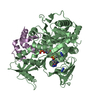 7thqC 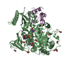 6o6eS S: Starting model for refinement C: citing same article ( |
|---|---|
| Similar structure data |
-Links
 PDBj
PDBj

- Assembly
| Deposited unit | 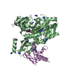
| ||||||||
|---|---|---|---|---|---|---|---|---|---|
| 1 |
| ||||||||
| Unit cell |
|
-Components
-Protein , 2 types, 2 molecules BE
| #1: Protein | Mass: 54823.832 Da / Num. of mol.: 1 Source method: isolated from a genetically manipulated source Source: (gene. exp.) Serratia sp. ATCC 39006 (bacteria) / Strain: ATCC 39006 / Gene: pigI / Production host: References: UniProt: Q5W263, L-proline-[L-prolyl-carrier protein] ligase |
|---|---|
| #2: Protein | Mass: 10689.031 Da / Num. of mol.: 1 Source method: isolated from a genetically manipulated source Source: (gene. exp.) Serratia sp. ATCC 39006 (bacteria) / Strain: ATCC 39006 / Gene: pigG / Production host: |
-Non-polymers , 5 types, 639 molecules 


| #3: Chemical | ChemComp-GOL /  Mass: 92.094 Da / Num. of mol.: 1 / Source method: obtained synthetically / Formula: C3H8O3 Mass: 92.094 Da / Num. of mol.: 1 / Source method: obtained synthetically / Formula: C3H8O3 | ||||||
|---|---|---|---|---|---|---|---|
| #4: Chemical | ChemComp-AZI /  Mass: 42.020 Da / Num. of mol.: 16 / Source method: obtained synthetically / Formula: N3 Mass: 42.020 Da / Num. of mol.: 16 / Source method: obtained synthetically / Formula: N3#5: Chemical | ChemComp-MG / |  Mass: 24.305 Da / Num. of mol.: 1 / Source method: obtained synthetically / Formula: Mg Mass: 24.305 Da / Num. of mol.: 1 / Source method: obtained synthetically / Formula: Mg#6: Chemical | ChemComp-I5M / |  Mass: 765.796 Da / Num. of mol.: 1 / Source method: obtained synthetically / Formula: C27H44N9O11PS2 / Feature type: SUBJECT OF INVESTIGATION Mass: 765.796 Da / Num. of mol.: 1 / Source method: obtained synthetically / Formula: C27H44N9O11PS2 / Feature type: SUBJECT OF INVESTIGATION#7: Water | ChemComp-HOH / | Mass: 18.015 Da / Num. of mol.: 620 / Source method: isolated from a natural source / Formula: H2O |
-Details
| Has ligand of interest | Y |
|---|---|
| Has protein modification | Y |
-Experimental details
-Experiment
| Experiment | Method: X-RAY DIFFRACTION / Number of used crystals: 1 |
|---|
- Sample preparation
| Crystal | Density Matthews: 2.24 Å3/Da / Density % sol: 45.11 % |
|---|---|
| Crystal grow | Temperature: 298 K / Method: vapor diffusion, hanging drop / pH: 6.23 Details: 0.3 M MgCl2, 24.57% PEG 3350, and 0.1 M BIS-TRIS pH 6.23 |
-Data collection
| Diffraction | Mean temperature: 110 K / Serial crystal experiment: N |
|---|---|
| Diffraction source | Source: SYNCHROTRON / Site: ALS / Beamline: 8.3.1 / Wavelength: 1.12 Å |
| Detector | Type: DECTRIS PILATUS3 S 6M / Detector: PIXEL / Date: Aug 16, 2019 |
| Radiation | Protocol: SINGLE WAVELENGTH / Monochromatic (M) / Laue (L): M / Scattering type: x-ray |
| Radiation wavelength | Wavelength: 1.12 Å / Relative weight: 1 |
| Reflection | Resolution: 1.6→62.55 Å / Num. obs: 510902 / % possible obs: 99.92 % / Redundancy: 6.8 % / CC1/2: 0.998 / CC star: 1 / Rmerge(I) obs: 0.1141 / Rpim(I) all: 0.04718 / Rrim(I) all: 0.1237 / Net I/σ(I): 12.66 |
| Reflection shell | Resolution: 1.6→1.662 Å / Redundancy: 6.2 % / Rmerge(I) obs: 1.045 / Mean I/σ(I) obs: 1.62 / Num. unique obs: 7504 / CC1/2: 0.603 / CC star: 0.867 / Rpim(I) all: 0.4542 / Rrim(I) all: 1.142 / % possible all: 99.93 |
- Processing
| Software |
| ||||||||||||||||||||||||||||||||||||||||||||||||||||||||||||
|---|---|---|---|---|---|---|---|---|---|---|---|---|---|---|---|---|---|---|---|---|---|---|---|---|---|---|---|---|---|---|---|---|---|---|---|---|---|---|---|---|---|---|---|---|---|---|---|---|---|---|---|---|---|---|---|---|---|---|---|---|---|
| Refinement | Method to determine structure: MOLECULAR REPLACEMENT Starting model: 6o6e Resolution: 1.6→62.55 Å / Cor.coef. Fo:Fc: 0.972 / Cor.coef. Fo:Fc free: 0.956 / SU B: 1.763 / SU ML: 0.06 / Cross valid method: THROUGHOUT / σ(F): 0 / ESU R: 0.077 / ESU R Free: 0.08 / Stereochemistry target values: MAXIMUM LIKELIHOOD Details: HYDROGENS HAVE BEEN ADDED IN THE RIDING POSITIONS U VALUES : REFINED INDIVIDUALLY
| ||||||||||||||||||||||||||||||||||||||||||||||||||||||||||||
| Solvent computation | Ion probe radii: 0.8 Å / Shrinkage radii: 0.8 Å / VDW probe radii: 1.2 Å / Solvent model: MASK | ||||||||||||||||||||||||||||||||||||||||||||||||||||||||||||
| Displacement parameters | Biso max: 73.1 Å2 / Biso mean: 17.858 Å2 / Biso min: 8.29 Å2
| ||||||||||||||||||||||||||||||||||||||||||||||||||||||||||||
| Refinement step | Cycle: final / Resolution: 1.6→62.55 Å
| ||||||||||||||||||||||||||||||||||||||||||||||||||||||||||||
| Refine LS restraints |
| ||||||||||||||||||||||||||||||||||||||||||||||||||||||||||||
| LS refinement shell | Resolution: 1.605→1.646 Å / Rfactor Rfree error: 0 / Total num. of bins used: 20
|
