 Movie
Movie Controller

[English] 日本語
 Yorodumi
- PDB-7tfi: Atomic model of the S. cerevisiae clamp-clamp loader complex PCNA... -
+ Open data

- Basic information
| Entry | Database: PDB / ID: 7tfi | |||||||||||||||||||||
|---|---|---|---|---|---|---|---|---|---|---|---|---|---|---|---|---|---|---|---|---|---|---|
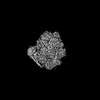
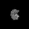
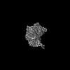
| Title | Atomic model of the S. cerevisiae clamp-clamp loader complex PCNA-RFC bound to DNA with an open clamp | |||||||||||||||||||||
 Components Components |
| |||||||||||||||||||||
 Keywords Keywords | DNA BINDING PROTEIN/DNA / DNA replication / DNA damage repair / RFC loader / PCNA clamp / DNA polymerase processivity factor / DNA binding protein-DNA complex | |||||||||||||||||||||
| Function / homology |  Function and homology information Function and homology informationpositive regulation of DNA metabolic process / Mismatch repair (MMR) directed by MSH2:MSH6 (MutSalpha) / meiotic mismatch repair / Rad17 RFC-like complex / : / Processive synthesis on the lagging strand / Removal of the Flap Intermediate / Elg1 RFC-like complex / DNA replication factor C complex / Ctf18 RFC-like complex ...positive regulation of DNA metabolic process / Mismatch repair (MMR) directed by MSH2:MSH6 (MutSalpha) / meiotic mismatch repair / Rad17 RFC-like complex / : / Processive synthesis on the lagging strand / Removal of the Flap Intermediate / Elg1 RFC-like complex / DNA replication factor C complex / Ctf18 RFC-like complex / : / Polymerase switching / maintenance of DNA trinucleotide repeats / DNA clamp loader activity / SUMOylation of DNA replication proteins / Translesion synthesis by REV1 / : / : / : / DNA replication checkpoint signaling / establishment of mitotic sister chromatid cohesion / PCNA complex / : / Activation of ATR in response to replication stress / lagging strand elongation / DNA damage tolerance / silent mating-type cassette heterochromatin formation / sister chromatid cohesion / mitotic sister chromatid cohesion / error-free translesion synthesis / DNA polymerase processivity factor activity / leading strand elongation / Gap-filling DNA repair synthesis and ligation in TC-NER / Dual incision in TC-NER / translesion synthesis / subtelomeric heterochromatin formation / mismatch repair / DNA damage checkpoint signaling / positive regulation of DNA repair / positive regulation of DNA replication / replication fork / nucleotide-excision repair / DNA-templated DNA replication / mitotic cell cycle / chromosome, telomeric region / cell division / DNA repair / ATP hydrolysis activity / DNA binding / ATP binding / identical protein binding / nucleus / cytosol Similarity search - Function | |||||||||||||||||||||
| Biological species |  synthetic construct (others) | |||||||||||||||||||||
| Method | ELECTRON MICROSCOPY / single particle reconstruction / cryo EM / Resolution: 3.41 Å | |||||||||||||||||||||
 Authors Authors | Zheng, F. / Georgescu, R. / Yao, Y.N. / O'Donnell, M.E. / Li, H. | |||||||||||||||||||||
| Funding support |  United States, 3items United States, 3items
| |||||||||||||||||||||
 Citation Citation | Journal: Elife / Year: 2022 Title: Cryo-EM structures reveal that RFC recognizes both the 3'- and 5'-DNA ends to load PCNA onto gaps for DNA repair. Authors: Fengwei Zheng / Roxana Georgescu / Nina Y Yao / Huilin Li / Michael E O'Donnell / Abstract: RFC uses ATP to assemble PCNA onto primed sites for replicative DNA polymerases δ and ε. The RFC pentamer forms a central chamber that binds 3' ss/ds DNA junctions to load PCNA onto DNA during ...RFC uses ATP to assemble PCNA onto primed sites for replicative DNA polymerases δ and ε. The RFC pentamer forms a central chamber that binds 3' ss/ds DNA junctions to load PCNA onto DNA during replication. We show here five structures that identify a second DNA binding site in RFC that binds a 5' duplex. This 5' DNA site is located between the N-terminal BRCT domain and AAA+ module of the large Rfc1 subunit. Our structures reveal ideal binding to a 7-nt gap, which includes 2 bp unwound by the clamp loader. Biochemical studies show enhanced binding to 5 and 10 nt gaps, consistent with the structural results. Because both 3' and 5' ends are present at a ssDNA gap, we propose that the 5' site facilitates RFC's PCNA loading activity at a DNA damage-induced gap to recruit gap-filling polymerases. These findings are consistent with genetic studies showing that base excision repair of gaps greater than 1 base requires PCNA and involves the 5' DNA binding domain of Rfc1. We further observe that a 5' end facilitates PCNA loading at an RPA coated 30-nt gap, suggesting a potential role of the RFC 5'-DNA site in lagging strand DNA synthesis. | |||||||||||||||||||||
| History |
|
- Structure visualization
| Structure viewer | Molecule: MolmilJmol/JSmol |
|---|
- Downloads & links
-Download
| PDBx/mmCIF format | 7tfi.cif.gz | 476.9 KB | Display | PDBx/mmCIF format |
|---|---|---|---|---|
| PDB format | pdb7tfi.ent.gz | 378 KB | Display | PDB format |
| PDBx/mmJSON format | 7tfi.json.gz | Tree view | PDBx/mmJSON format | |
| Others |  Other downloads Other downloads |
-Validation report
| Arichive directory | https://data.pdbj.org/pub/pdb/validation_reports/tf/7tfiftp://data.pdbj.org/pub/pdb/validation_reports/tf/7tfi | HTTPS FTP |
|---|
-Related structure data
| Related structure data | 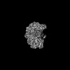 25873MC  7tfhC  7tfjC  7tfkC  7tflC M: map data used to model this data C: citing same article ( |
|---|---|
| Similar structure data |
-Links
 PDBj
PDBj


- Assembly
| Deposited unit | 
|
|---|---|
| 1 |
|
-Components
-Replication factor C subunit ... , 5 types, 5 molecules ABCDE
| #1: Protein | Mass: 95048.195 Da / Num. of mol.: 1 Source method: isolated from a genetically manipulated source Source: (gene. exp.) Gene: RFC1, CDC44, YOR217W, YOR50-7 / Production host:  |
|---|---|
| #2: Protein | Mass: 36201.039 Da / Num. of mol.: 1 Source method: isolated from a genetically manipulated source Source: (gene. exp.) Gene: RFC4, YOL094C, O0923 / Production host: |
| #3: Protein | Mass: 38254.543 Da / Num. of mol.: 1 Source method: isolated from a genetically manipulated source Source: (gene. exp.) Gene: RFC3, YNL290W, N0533 / Production host: |
| #4: Protein | Mass: 39794.473 Da / Num. of mol.: 1 Source method: isolated from a genetically manipulated source Source: (gene. exp.) Gene: RFC2, YJR068W, J1808 / Production host: |
| #5: Protein | Mass: 39993.582 Da / Num. of mol.: 1 Source method: isolated from a genetically manipulated source Source: (gene. exp.) Gene: RFC5, YBR087W, YBR0810 / Production host: |
-Protein , 1 types, 3 molecules FGH
| #6: Protein | Mass: 29383.570 Da / Num. of mol.: 3 Source method: isolated from a genetically manipulated source Source: (gene. exp.) Gene: POL30, YBR088C, YBR0811 / Production host: |
|---|
-DNA chain , 2 types, 2 molecules IJ
| #7: DNA chain | Mass: 12214.818 Da / Num. of mol.: 1 / Source method: obtained synthetically / Source: (synth.) synthetic construct (others) |
|---|---|
| #8: DNA chain | Mass: 6136.008 Da / Num. of mol.: 1 / Source method: obtained synthetically / Source: (synth.) synthetic construct (others) |
-Non-polymers , 3 types, 9 molecules 


| #9: Chemical | ChemComp-AGS /  Mass: 523.247 Da / Num. of mol.: 4 / Source method: obtained synthetically / Formula: C10H16N5O12P3S / Comment: ATP-gamma-S, energy-carrying molecule analogue*YM Mass: 523.247 Da / Num. of mol.: 4 / Source method: obtained synthetically / Formula: C10H16N5O12P3S / Comment: ATP-gamma-S, energy-carrying molecule analogue*YM#10: Chemical | ChemComp-MG /  Mass: 24.305 Da / Num. of mol.: 4 / Source method: obtained synthetically / Formula: Mg Mass: 24.305 Da / Num. of mol.: 4 / Source method: obtained synthetically / Formula: Mg#11: Chemical | ChemComp-ADP / |  Mass: 427.201 Da / Num. of mol.: 1 / Source method: obtained synthetically / Formula: C10H15N5O10P2 / Comment: ADP, energy-carrying molecule*YM Mass: 427.201 Da / Num. of mol.: 1 / Source method: obtained synthetically / Formula: C10H15N5O10P2 / Comment: ADP, energy-carrying molecule*YM |
|---|
-Details
| Has ligand of interest | Y |
|---|---|
| Has protein modification | Y |
-Experimental details
-Experiment
| Experiment | Method: ELECTRON MICROSCOPY |
|---|---|
| EM experiment | Aggregation state: PARTICLE / 3D reconstruction method: single particle reconstruction |
- Sample preparation
| Component |
| ||||||||||||||||||||||||||||
|---|---|---|---|---|---|---|---|---|---|---|---|---|---|---|---|---|---|---|---|---|---|---|---|---|---|---|---|---|---|
| Source (natural) |
| ||||||||||||||||||||||||||||
| Source (recombinant) | Organism: | ||||||||||||||||||||||||||||
| Buffer solution | pH: 7.5 | ||||||||||||||||||||||||||||
| Specimen | Embedding applied: NO / Shadowing applied: NO / Staining applied: NO / Vitrification applied: YES | ||||||||||||||||||||||||||||
| Vitrification | Cryogen name: ETHANE |
- Electron microscopy imaging
| Experimental equipment |  Model: Titan Krios / Image courtesy: FEI Company |
|---|---|
| Microscopy | Model: FEI TITAN KRIOS |
| Electron gun | Electron source:  FIELD EMISSION GUN / Accelerating voltage: 300 kV / Illumination mode: FLOOD BEAM FIELD EMISSION GUN / Accelerating voltage: 300 kV / Illumination mode: FLOOD BEAM |
| Electron lens | Mode: BRIGHT FIELD / Nominal defocus max: 1900 nm / Nominal defocus min: 1300 nm |
| Image recording | Electron dose: 65 e/Å2 / Film or detector model: GATAN K3 (6k x 4k) |
- Processing
| Software | Name: PHENIX / Version: 1.19.2_4158: / Classification: refinement | ||||||||||||||||||||||||
|---|---|---|---|---|---|---|---|---|---|---|---|---|---|---|---|---|---|---|---|---|---|---|---|---|---|
| EM software | Name: PHENIX / Category: model refinement | ||||||||||||||||||||||||
| CTF correction | Type: NONE | ||||||||||||||||||||||||
| 3D reconstruction | Resolution: 3.41 Å / Resolution method: FSC 0.143 CUT-OFF / Num. of particles: 118384 / Symmetry type: POINT | ||||||||||||||||||||||||
| Refine LS restraints |
|
