 Movie
Movie Controller


+ Open data

- Basic information
| Entry | Database: PDB / ID: 7tbx | ||||||
|---|---|---|---|---|---|---|---|
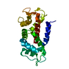
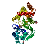
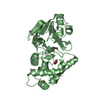
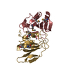
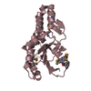
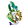
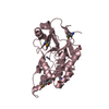
| Title | Crystal structure of D179Y KPC-2 beta-lactamase | ||||||
 Components Components | Carbapenem-hydrolyzing beta-lactamase KPC | ||||||
 Keywords Keywords | HYDROLASE / beta-lactamase / antibiotic resistance | ||||||
| Function / homology | Beta-lactamase class A, catalytic domain / Beta-lactamase enzyme family / Beta-lactamase, class-A / beta-lactam antibiotic catabolic process / Beta-lactamase/transpeptidase-like / beta-lactamase activity / beta-lactamase / response to antibiotic / Carbapenem-hydrolyzing beta-lactamase KPC Function and homology information Function and homology information | ||||||
| Biological species |  Klebsiella pneumoniae (bacteria) Klebsiella pneumoniae (bacteria) | ||||||
| Method |  X-RAY DIFFRACTION / SYNCHROTRON / MOLECULAR REPLACEMENT / Resolution: 3.16 Å X-RAY DIFFRACTION / SYNCHROTRON / MOLECULAR REPLACEMENT / Resolution: 3.16 Å | ||||||
 Authors Authors | van den Akker, F. / Alsenani, T. | ||||||
| Funding support |  United States, 1items United States, 1items
| ||||||
 Citation Citation | Journal: Antimicrob.Agents Chemother. / Year: 2022 Title: Structural Characterization of the D179N and D179Y Variants of KPC-2 beta-Lactamase: Omega-Loop Destabilization as a Mechanism of Resistance to Ceftazidime-Avibactam. Authors: Alsenani, T.A. / Viviani, S.L. / Kumar, V. / Taracila, M.A. / Bethel, C.R. / Barnes, M.D. / Papp-Wallace, K.M. / Shields, R.K. / Nguyen, M.H. / Clancy, C.J. / Bonomo, R.A. / van den Akker, F. | ||||||
| History |
|
- Structure visualization
| Structure viewer | Molecule: MolmilJmol/JSmol |
|---|
- Downloads & links
-Download
| PDBx/mmCIF format | 7tbx.cif.gz | 98.4 KB | Display | PDBx/mmCIF format |
|---|---|---|---|---|
| PDB format | pdb7tbx.ent.gz | 73 KB | Display | PDB format |
| PDBx/mmJSON format | 7tbx.json.gz | Tree view | PDBx/mmJSON format | |
| Others |  Other downloads Other downloads |
-Validation report
| Summary document | 7tbx_validation.pdf.gz | 435.2 KB | Display | wwPDB validaton report |
|---|---|---|---|---|
| Full document | 7tbx_full_validation.pdf.gz | 440.6 KB | Display | |
| Data in XML | 7tbx_validation.xml.gz | 17.5 KB | Display | |
| Data in CIF | 7tbx_validation.cif.gz | 22.6 KB | Display | |
| Arichive directory | https://data.pdbj.org/pub/pdb/validation_reports/tb/7tbxftp://data.pdbj.org/pub/pdb/validation_reports/tb/7tbx | HTTPS FTP |
-Related structure data
| Related structure data | 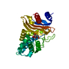 7tb7C 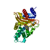 7tc1C 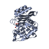 2ov5S C: citing same article ( S: Starting model for refinement |
|---|---|
| Similar structure data |


-Links
 PDBj
PDBj


- Assembly
| Deposited unit | 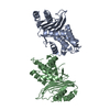
| ||||||||
|---|---|---|---|---|---|---|---|---|---|
| 1 | 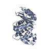
| ||||||||
| 2 | 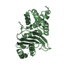
| ||||||||
| Unit cell |
|
-Components
| #1: Protein | Mass: 28325.930 Da / Num. of mol.: 2 / Mutation: D179Y Source method: isolated from a genetically manipulated source Source: (gene. exp.) Klebsiella pneumoniae (bacteria) / Gene: bla, kpc, kpc1 / Production host: |
|---|
-Experimental details
-Experiment
| Experiment | Method: X-RAY DIFFRACTION / Number of used crystals: 1 |
|---|
- Sample preparation
| Crystal | Density Matthews: 3.48 Å3/Da / Density % sol: 64.66 % |
|---|---|
| Crystal grow | Temperature: 298 K / Method: vapor diffusion, sitting drop / pH: 4.6 Details: 1 M ammonium citrate dibasic, 0.1 M sodium acetate trihydrate pH 4.6 |
-Data collection
| Diffraction | Mean temperature: 100 K / Serial crystal experiment: N | ||||||||||||||||||||||||||||||
|---|---|---|---|---|---|---|---|---|---|---|---|---|---|---|---|---|---|---|---|---|---|---|---|---|---|---|---|---|---|---|---|
| Diffraction source | Source: SYNCHROTRON / Site: NSLS-II / Beamline: 17-ID-2 / Wavelength: 0.9201 Å | ||||||||||||||||||||||||||||||
| Detector | Type: DECTRIS EIGER X 16M / Detector: PIXEL / Date: Mar 22, 2021 | ||||||||||||||||||||||||||||||
| Radiation | Protocol: SINGLE WAVELENGTH / Monochromatic (M) / Laue (L): M / Scattering type: x-ray | ||||||||||||||||||||||||||||||
| Radiation wavelength | Wavelength: 0.9201 Å / Relative weight: 1 | ||||||||||||||||||||||||||||||
| Reflection | Resolution: 3.13→27.32 Å / Num. obs: 13416 / % possible obs: 97.4 % / Redundancy: 5.1 % / Biso Wilson estimate: 119.87 Å2 / CC1/2: 0.992 / Rmerge(I) obs: 0.195 / Rpim(I) all: 0.095 / Rrim(I) all: 0.217 / Net I/σ(I): 4.5 / Num. measured all: 69003 | ||||||||||||||||||||||||||||||
| Reflection shell | Diffraction-ID: 1
|
- Processing
| Software |
| ||||||||||||||||||||||||||||||||||||||||||
|---|---|---|---|---|---|---|---|---|---|---|---|---|---|---|---|---|---|---|---|---|---|---|---|---|---|---|---|---|---|---|---|---|---|---|---|---|---|---|---|---|---|---|---|
| Refinement | Method to determine structure: MOLECULAR REPLACEMENT Starting model: 2OV5 Resolution: 3.16→27.32 Å / SU ML: 0.92 / Cross valid method: THROUGHOUT / σ(F): 1.35 / Phase error: 44.78 / Stereochemistry target values: ML
| ||||||||||||||||||||||||||||||||||||||||||
| Solvent computation | Shrinkage radii: 0.9 Å / VDW probe radii: 1.11 Å / Solvent model: FLAT BULK SOLVENT MODEL | ||||||||||||||||||||||||||||||||||||||||||
| Displacement parameters | Biso max: 291.24 Å2 / Biso mean: 129.4946 Å2 / Biso min: 65.41 Å2 | ||||||||||||||||||||||||||||||||||||||||||
| Refinement step | Cycle: final / Resolution: 3.16→27.32 Å
| ||||||||||||||||||||||||||||||||||||||||||
| LS refinement shell | Refine-ID: X-RAY DIFFRACTION / Rfactor Rfree error: 0 / Total num. of bins used: 5
|
