 Movie
Movie Controller

[English] 日本語
 Yorodumi
- PDB-7ta0: Human Ornithine Aminotransferase (hOAT) soaked with 5-aminovaleri... -
+ Open data

- Basic information
| Entry | Database: PDB / ID: 7ta0 | ||||||
|---|---|---|---|---|---|---|---|
| Title | Human Ornithine Aminotransferase (hOAT) soaked with 5-aminovaleric acid | ||||||
 Components Components | Ornithine aminotransferase, mitochondrial | ||||||
 Keywords Keywords | TRANSFERASE / Human Ornithine Aminotransferase / hOAT / OAT / PLP / 5-aminovaleric acid / soaking | ||||||
| Function / homology |  Function and homology information Function and homology information: / ornithine aminotransferase / L-ornithine transaminase activity / : / L-proline biosynthetic process / Glutamate and glutamine metabolism / visual perception / pyridoxal phosphate binding / mitochondrial matrix / mitochondrion ...: / ornithine aminotransferase / L-ornithine transaminase activity / : / L-proline biosynthetic process / Glutamate and glutamine metabolism / visual perception / pyridoxal phosphate binding / mitochondrial matrix / mitochondrion / nucleoplasm / identical protein binding / cytoplasm Similarity search - Function | ||||||
| Biological species |  Homo sapiens (human) Homo sapiens (human) | ||||||
| Method |  X-RAY DIFFRACTION / SYNCHROTRON / MOLECULAR REPLACEMENT / Resolution: 2.33 Å X-RAY DIFFRACTION / SYNCHROTRON / MOLECULAR REPLACEMENT / Resolution: 2.33 Å | ||||||
 Authors Authors | Butrin, A. / Liu, D. | ||||||
| Funding support |  United States, 1items United States, 1items
| ||||||
 Citation Citation | Journal: J.Biol.Chem. / Year: 2022 Title: Determination of the pH dependence, substrate specificity, and turnovers of alternative substrates for human ornithine aminotransferase. Authors: Butrin, A. / Butrin, A. / Wawrzak, Z. / Moran, G.R. / Liu, D. | ||||||
| History |
|
- Structure visualization
| Structure viewer | Molecule: MolmilJmol/JSmol |
|---|
- Downloads & links
-Download
| PDBx/mmCIF format | 7ta0.cif.gz | 248.6 KB | Display | PDBx/mmCIF format |
|---|---|---|---|---|
| PDB format | pdb7ta0.ent.gz | 200.7 KB | Display | PDB format |
| PDBx/mmJSON format | 7ta0.json.gz | Tree view | PDBx/mmJSON format | |
| Others |  Other downloads Other downloads |
-Validation report
| Arichive directory | https://data.pdbj.org/pub/pdb/validation_reports/ta/7ta0ftp://data.pdbj.org/pub/pdb/validation_reports/ta/7ta0 | HTTPS FTP |
|---|
-Related structure data
| Related structure data |  7t9zC  7ta1C 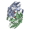 1oatS S: Starting model for refinement C: citing same article ( |
|---|---|
| Similar structure data |
-Links
 PDBj
PDBj- Assembly
| Deposited unit | 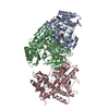
| ||||||||||
|---|---|---|---|---|---|---|---|---|---|---|---|
| 1 | 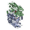
| ||||||||||
| 2 | 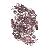
| ||||||||||
| Unit cell |
| ||||||||||
| Components on special symmetry positions |
|
-Components
| #1: Protein | Mass: 48593.668 Da / Num. of mol.: 3 Source method: isolated from a genetically manipulated source Source: (gene. exp.) Homo sapiens (human) / Gene: OAT / Production host:  #2: Chemical | 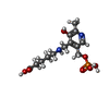  Mass: 348.289 Da / Num. of mol.: 3 / Source method: obtained synthetically / Formula: C13H21N2O7P Mass: 348.289 Da / Num. of mol.: 3 / Source method: obtained synthetically / Formula: C13H21N2O7P#3: Chemical |   Mass: 94.971 Da / Num. of mol.: 2 / Source method: obtained synthetically / Formula: PO4 Mass: 94.971 Da / Num. of mol.: 2 / Source method: obtained synthetically / Formula: PO4#4: Water | ChemComp-HOH / |  Mass: 18.015 Da / Num. of mol.: 325 / Source method: isolated from a natural source / Formula: H2O Mass: 18.015 Da / Num. of mol.: 325 / Source method: isolated from a natural source / Formula: H2OHas ligand of interest | Y | Has protein modification | Y | |
|---|
-Experimental details
-Experiment
| Experiment | Method: X-RAY DIFFRACTION / Number of used crystals: 1 |
|---|
- Sample preparation
| Crystal | Density Matthews: 2.42 Å3/Da / Density % sol: 49.19 % |
|---|---|
| Crystal grow | Temperature: 293 K / Method: vapor diffusion, hanging drop / pH: 7.8 Details: Once hOAT was purified, it was transferred to a 10 kDa centrifugal filter tube and concentrated to ~6 mg/mL. The holoenzyme crystals were first grown via a hanging drop vapor diffusion ...Details: Once hOAT was purified, it was transferred to a 10 kDa centrifugal filter tube and concentrated to ~6 mg/mL. The holoenzyme crystals were first grown via a hanging drop vapor diffusion method. Each drop contained 2 uL of protein and 2 uL of well solution. The best crystallization condition contained 8% PEG 6000, 100 mM NaCl, 5% glycerol, and 50 mM Tricine pH 7.8. Once holoenzyme crystals reached their maximum size within seven days, 1 uL of 5-aminovaleric acid was added to the drop with crystals. The crystals were soaked for different time periods from 3 to 59 minutes. After soaking, crystals were transferred into a cryoprotective solution (well solution supplemented with 30% glycerol), and then flash-frozen in liquid nitrogen. |
-Data collection
| Diffraction | Mean temperature: 100 K / Serial crystal experiment: N |
|---|---|
| Diffraction source | Source: SYNCHROTRON / Site: APS / Beamline: 21-ID-D / Wavelength: 1.127 Å |
| Detector | Type: DECTRIS EIGER X 9M / Detector: PIXEL / Date: Jul 3, 2021 |
| Radiation | Protocol: SINGLE WAVELENGTH / Monochromatic (M) / Laue (L): M / Scattering type: x-ray |
| Radiation wavelength | Wavelength: 1.127 Å / Relative weight: 1 |
| Reflection | Resolution: 2.326→99.512 Å / Num. obs: 61264 / % possible obs: 99.9 % / Redundancy: 15.3 % / Biso Wilson estimate: 41.16 Å2 / CC1/2: 0.994 / Net I/σ(I): 7 |
| Reflection shell | Resolution: 2.326→2.366 Å / Num. unique obs: 2995 / CC1/2: 0.33 / % possible all: 100 |
- Processing
| Software |
| |||||||||||||||||||||||||||||||||||||||||||||||||||||||||||||||||||||||||||||||||||||||||||||||||||||||||||||||||||||||||||||||||||||||||||||||||||||||||||||||||
|---|---|---|---|---|---|---|---|---|---|---|---|---|---|---|---|---|---|---|---|---|---|---|---|---|---|---|---|---|---|---|---|---|---|---|---|---|---|---|---|---|---|---|---|---|---|---|---|---|---|---|---|---|---|---|---|---|---|---|---|---|---|---|---|---|---|---|---|---|---|---|---|---|---|---|---|---|---|---|---|---|---|---|---|---|---|---|---|---|---|---|---|---|---|---|---|---|---|---|---|---|---|---|---|---|---|---|---|---|---|---|---|---|---|---|---|---|---|---|---|---|---|---|---|---|---|---|---|---|---|---|---|---|---|---|---|---|---|---|---|---|---|---|---|---|---|---|---|---|---|---|---|---|---|---|---|---|---|---|---|---|---|---|
| Refinement | Method to determine structure: MOLECULAR REPLACEMENT Starting model: 1OAT Resolution: 2.33→99.51 Å / SU ML: 0.332 / Cross valid method: FREE R-VALUE / σ(F): 1.33 / Phase error: 26.964 Stereochemistry target values: GEOSTD + MONOMER LIBRARY + CDL V1.2
| |||||||||||||||||||||||||||||||||||||||||||||||||||||||||||||||||||||||||||||||||||||||||||||||||||||||||||||||||||||||||||||||||||||||||||||||||||||||||||||||||
| Solvent computation | Shrinkage radii: 0.9 Å / VDW probe radii: 1.11 Å / Solvent model: FLAT BULK SOLVENT MODEL | |||||||||||||||||||||||||||||||||||||||||||||||||||||||||||||||||||||||||||||||||||||||||||||||||||||||||||||||||||||||||||||||||||||||||||||||||||||||||||||||||
| Displacement parameters | Biso mean: 45.8 Å2 | |||||||||||||||||||||||||||||||||||||||||||||||||||||||||||||||||||||||||||||||||||||||||||||||||||||||||||||||||||||||||||||||||||||||||||||||||||||||||||||||||
| Refinement step | Cycle: LAST / Resolution: 2.33→99.51 Å
| |||||||||||||||||||||||||||||||||||||||||||||||||||||||||||||||||||||||||||||||||||||||||||||||||||||||||||||||||||||||||||||||||||||||||||||||||||||||||||||||||
| Refine LS restraints |
| |||||||||||||||||||||||||||||||||||||||||||||||||||||||||||||||||||||||||||||||||||||||||||||||||||||||||||||||||||||||||||||||||||||||||||||||||||||||||||||||||
| LS refinement shell |
|
