 Movie
Movie Controller


+ Open data

- Basic information
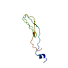
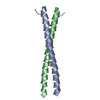
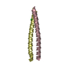
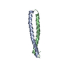
| Entry | Database: PDB / ID: 7t6y | |||||||||||||||||||||||||||||||
|---|---|---|---|---|---|---|---|---|---|---|---|---|---|---|---|---|---|---|---|---|---|---|---|---|---|---|---|---|---|---|---|---|
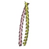
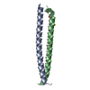
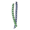
| Title | d((CGA)5TGA) parallel-stranded homo-duplex | |||||||||||||||||||||||||||||||
 Components Components | DNA (5'-D(* Keywords KeywordsDNA / triplet repeat DNA / parallel-stranded duplex / non-canonical DNA / d(CGA) | Function / homology | : / DNA / DNA (> 10) |  Function and homology information Function and homology informationBiological species | synthetic construct (others) | Method |  X-RAY DIFFRACTION / SYNCHROTRON / MOLECULAR REPLACEMENT / Resolution: 2.3 Å X-RAY DIFFRACTION / SYNCHROTRON / MOLECULAR REPLACEMENT / Resolution: 2.3 Å  Authors AuthorsLuteran, E.M. / Paukstelis, P.J. | Funding support | 1items |
 CitationJournal: Acta Crystallogr D Struct Biol / Year: 2022 CitationJournal: Acta Crystallogr D Struct Biol / Year: 2022Title: The parallel-stranded d(CGA) duplex is a highly predictable structural motif with two conformationally distinct strands. Authors: Luteran, E.M. / Paukstelis, P.J. History |
|
- Structure visualization
| Structure viewer | Molecule: MolmilJmol/JSmol |
|---|
- Downloads & links
-Download
| PDBx/mmCIF format | 7t6y.cif.gz | 37.1 KB | Display | PDBx/mmCIF format |
|---|---|---|---|---|
| PDB format | pdb7t6y.ent.gz | 22.8 KB | Display | PDB format |
| PDBx/mmJSON format | 7t6y.json.gz | Tree view | PDBx/mmJSON format | |
| Others |  Other downloads Other downloads |
-Validation report
| Arichive directory | https://data.pdbj.org/pub/pdb/validation_reports/t6/7t6yftp://data.pdbj.org/pub/pdb/validation_reports/t6/7t6y | HTTPS FTP |
|---|
-Related structure data
| Related structure data |  7sb8C  1ixjS C: citing same article ( S: Starting model for refinement |
|---|---|
| Similar structure data |

-Links
 PDBj
PDBj


- Assembly
| Deposited unit | 
| ||||||||
|---|---|---|---|---|---|---|---|---|---|
| 1 |
| ||||||||
| Unit cell |
| ||||||||
| Components on special symmetry positions |
|
-Components
| #1: DNA chain | Mass: 5559.620 Da / Num. of mol.: 2 Source method: isolated from a genetically manipulated source Source: (gene. exp.) synthetic construct (others) / Production host: synthetic construct (others) #2: Chemical | ChemComp-BA /   Mass: 137.327 Da / Num. of mol.: 7 / Source method: obtained synthetically / Formula: Ba Mass: 137.327 Da / Num. of mol.: 7 / Source method: obtained synthetically / Formula: Ba#3: Water | ChemComp-HOH / |  Mass: 18.015 Da / Num. of mol.: 119 / Source method: isolated from a natural source / Formula: H2O Mass: 18.015 Da / Num. of mol.: 119 / Source method: isolated from a natural source / Formula: H2OHas ligand of interest | N | |
|---|
-Experimental details
-Experiment
| Experiment | Method: X-RAY DIFFRACTION / Number of used crystals: 1 |
|---|
- Sample preparation
| Crystal | Density Matthews: 1.98 Å3/Da / Density % sol: 37.95 % |
|---|---|
| Crystal grow | Temperature: 295 K / Method: vapor diffusion, sitting drop / pH: 5.5 Details: 20% MPD, 120 mM barium chloride, 30 mM sodium cacodylate, equilibrated against 30% MPD |
-Data collection
| Diffraction | Mean temperature: 100 K / Serial crystal experiment: N |
|---|---|
| Diffraction source | Source: SYNCHROTRON / Site: APS  / Beamline: 24-ID-E / Wavelength: 0.979 Å / Beamline: 24-ID-E / Wavelength: 0.979 Å |
| Detector | Type: DECTRIS EIGER X 16M / Detector: PIXEL / Date: Nov 24, 2019 |
| Radiation | Protocol: SINGLE WAVELENGTH / Monochromatic (M) / Laue (L): M / Scattering type: x-ray |
| Radiation wavelength | Wavelength: 0.979 Å / Relative weight: 1 |
| Reflection | Resolution: 2.3→32.255 Å / Num. obs: 3487 / % possible obs: 87.2 % / Redundancy: 1.8 % / CC1/2: 0.94 / Rpim(I) all: 0.109 / Net I/σ(I): 5.7 |
| Reflection shell | Resolution: 2.3→2.38 Å / Mean I/σ(I) obs: 5 / Num. unique obs: 350 / CC1/2: 0.395 / Rpim(I) all: 0.288 |
- Processing
| Software |
| ||||||||||||||||||||||||
|---|---|---|---|---|---|---|---|---|---|---|---|---|---|---|---|---|---|---|---|---|---|---|---|---|---|
| Refinement | Method to determine structure: MOLECULAR REPLACEMENT Starting model: 1IXJ Resolution: 2.3→32.255 Å / SU ML: 0.19 / Cross valid method: THROUGHOUT / σ(F): 1.45 / Phase error: 19.5 / Stereochemistry target values: ML
| ||||||||||||||||||||||||
| Solvent computation | Shrinkage radii: 0.9 Å / VDW probe radii: 1.11 Å / Solvent model: FLAT BULK SOLVENT MODEL | ||||||||||||||||||||||||
| Displacement parameters | Biso max: 41.02 Å2 / Biso mean: 13.2239 Å2 / Biso min: 1.79 Å2 | ||||||||||||||||||||||||
| Refinement step | Cycle: final / Resolution: 2.3→32.255 Å
| ||||||||||||||||||||||||
| Refine LS restraints |
| ||||||||||||||||||||||||
| LS refinement shell | Refine-ID: X-RAY DIFFRACTION / Rfactor Rfree error: 0 / % reflection obs: 87 %
|
