 ムービー
ムービー コントローラー


+ データを開く

- 基本情報
基本情報
| 登録情報 | データベース: PDB / ID: 7t6f | ||||||||||||
|---|---|---|---|---|---|---|---|---|---|---|---|---|---|
| タイトル | Structure of active Janus Kinase (JAK) dimer complexed with cytokine receptor intracellular domain | ||||||||||||
 要素 要素 |
| ||||||||||||
 キーワード キーワード | SIGNALING PROTEIN / signaling complex / Janus Kinase / JAK / oncogenic mutation / gain-of-function mutation / cytokine receptor | ||||||||||||
| 機能・相同性 |  機能・相同性情報 機能・相同性情報interleukin-28 receptor complex / response to type III interferon / Interleukin-20 family signaling / mucosal immune response / positive regulation of cellular respiration / protein localization to cell-cell junction / interleukin-11-mediated signaling pathway / CCR5 chemokine receptor binding / interleukin-9-mediated signaling pathway / interleukin-4-mediated signaling pathway ...interleukin-28 receptor complex / response to type III interferon / Interleukin-20 family signaling / mucosal immune response / positive regulation of cellular respiration / protein localization to cell-cell junction / interleukin-11-mediated signaling pathway / CCR5 chemokine receptor binding / interleukin-9-mediated signaling pathway / interleukin-4-mediated signaling pathway / interleukin-10-mediated signaling pathway / interleukin-2-mediated signaling pathway / positive regulation of homotypic cell-cell adhesion / regulation of defense response to virus by host / interleukin-15-mediated signaling pathway / cytokine receptor activity / growth hormone receptor binding / extrinsic component of cytoplasmic side of plasma membrane / type I interferon-mediated signaling pathway / interleukin-6-mediated signaling pathway / positive regulation of sprouting angiogenesis / cell surface receptor signaling pathway via JAK-STAT / type II interferon-mediated signaling pathway / non-membrane spanning protein tyrosine kinase activity / non-specific protein-tyrosine kinase / positive regulation of protein localization to nucleus / protein phosphatase binding / defense response to virus / cytoskeleton / receptor complex / intracellular signal transduction / response to antibiotic / ubiquitin protein ligase binding / ATP binding / nucleus / cytoplasm 類似検索 - 分子機能 | ||||||||||||
| 生物種 |  | ||||||||||||
| 手法 | 電子顕微鏡法 / 単粒子再構成法 / クライオ電子顕微鏡法 / 解像度: 3.6 Å | ||||||||||||
 データ登録者 データ登録者 | Glassman, C.R. / Tsutsumi, N. / Jude, K.M. / Garcia, K.C. | ||||||||||||
| 資金援助 |  米国, 3件 米国, 3件
| ||||||||||||
 引用 引用 | ジャーナル: Science / 年: 2022 タイトル: Structure of a Janus kinase cytokine receptor complex reveals the basis for dimeric activation. 著者: Caleb R Glassman / Naotaka Tsutsumi / Robert A Saxton / Patrick J Lupardus / Kevin M Jude / K Christopher Garcia / 要旨: Cytokines signal through cell surface receptor dimers to initiate activation of intracellular Janus kinases (JAKs). We report the 3.6-angstrom-resolution cryo-electron microscopy structure of full- ...Cytokines signal through cell surface receptor dimers to initiate activation of intracellular Janus kinases (JAKs). We report the 3.6-angstrom-resolution cryo-electron microscopy structure of full-length JAK1 complexed with a cytokine receptor intracellular domain Box1 and Box2 regions captured as an activated homodimer bearing the valine→phenylalanine (VF) mutation prevalent in myeloproliferative neoplasms. The seven domains of JAK1 form an extended structural unit, the dimerization of which is mediated by close-packing of the pseudokinase (PK) domains from the monomeric subunits. The oncogenic VF mutation lies within the core of the JAK1 PK interdimer interface, enhancing packing complementarity to facilitate ligand-independent activation. The carboxy-terminal tyrosine kinase domains are poised for transactivation and to phosphorylate the receptor STAT (signal transducer and activator of transcription)-recruiting motifs projecting from the overhanging FERM (four-point-one, ezrin, radixin, moesin)-SH2 (Src homology 2)-domains. Mapping of constitutively active JAK mutants supports a two-step allosteric activation mechanism and reveals opportunities for selective therapeutic targeting of oncogenic JAK signaling. | ||||||||||||
| 履歴 |
|
- 構造の表示
構造の表示
| ムービー |
ムービービューア |
|---|---|
| 構造ビューア | 分子: MolmilJmol/JSmol |
- ダウンロードとリンク
-ダウンロード
| PDBx/mmCIF形式 | 7t6f.cif.gz | 394.5 KB | 表示 | PDBx/mmCIF形式 |
|---|---|---|---|---|
| PDB形式 | pdb7t6f.ent.gz | 311.8 KB | 表示 | PDB形式 |
| PDBx/mmJSON形式 | 7t6f.json.gz | ツリー表示 | PDBx/mmJSON形式 | |
| その他 |  その他のダウンロード その他のダウンロード |
-検証レポート
| 文書・要旨 | 7t6f_validation.pdf.gz | 1.7 MB | 表示 | wwPDB検証レポート |
|---|---|---|---|---|
| 文書・詳細版 | 7t6f_full_validation.pdf.gz | 1.7 MB | 表示 | |
| XML形式データ | 7t6f_validation.xml.gz | 70.6 KB | 表示 | |
| CIF形式データ | 7t6f_validation.cif.gz | 104.6 KB | 表示 | |
| アーカイブディレクトリ | https://data.pdbj.org/pub/pdb/validation_reports/t6/7t6fftp://data.pdbj.org/pub/pdb/validation_reports/t6/7t6f | HTTPS FTP |
-関連構造データ


-リンク
 PDBj
PDBj


- 集合体
集合体
| 登録構造単位 | 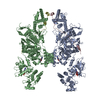
|
|---|---|
| 1 |
|
-要素
| #1: タンパク質 | 分子量: 136026.844 Da / 分子数: 2 / 変異: V657F / 由来タイプ: 組換発現 / 由来: (組換発現)  Trichoplusia ni (イラクサキンウワバ) Trichoplusia ni (イラクサキンウワバ)参照: UniProt: B1ASP2, non-specific protein-tyrosine kinase #2: タンパク質 | 分子量: 9895.373 Da / 分子数: 2 / 由来タイプ: 組換発現 / 由来: (組換発現) Trichoplusia ni (イラクサキンウワバ) / 参照: UniProt: Q8CGK5#3: 化合物 |   分子量: 267.241 Da / 分子数: 2 / 由来タイプ: 合成 / 式: C10H13N5O4 分子量: 267.241 Da / 分子数: 2 / 由来タイプ: 合成 / 式: C10H13N5O4#4: 化合物 |   分子量: 427.201 Da / 分子数: 2 / 由来タイプ: 合成 / 式: C10H15N5O10P2 / コメント: ADP, エネルギー貯蔵分子*YM 分子量: 427.201 Da / 分子数: 2 / 由来タイプ: 合成 / 式: C10H15N5O10P2 / コメント: ADP, エネルギー貯蔵分子*YM研究の焦点であるリガンドがあるか | N | |
|---|
-実験情報
-実験
| 実験 | 手法: 電子顕微鏡法 |
|---|---|
| EM実験 | 試料の集合状態: PARTICLE / 3次元再構成法: 単粒子再構成法 |
- 試料調製
試料調製
| 構成要素 | 名称: GCN4-zippered dimeric IFN-lambda intracellular domain bound to two Jak1s タイプ: COMPLEX 詳細: Co-expressed GST-fused GCN4-IFN-lambda and full-length Jak1 in T. ni. GST tagged was removed by 3C protease digestion. Entity ID: #1-#2 / 由来: RECOMBINANT | ||||||||||||||||||||||||
|---|---|---|---|---|---|---|---|---|---|---|---|---|---|---|---|---|---|---|---|---|---|---|---|---|---|
| 分子量 | 値: 0.29 MDa / 実験値: NO | ||||||||||||||||||||||||
| 由来(天然) | 生物種: | ||||||||||||||||||||||||
| 由来(組換発現) | 生物種: Trichoplusia ni (イラクサキンウワバ) | ||||||||||||||||||||||||
| 緩衝液 | pH: 8 / 詳細: additive: 0.01% w/v DDM | ||||||||||||||||||||||||
| 緩衝液成分 |
| ||||||||||||||||||||||||
| 試料 | 濃度: 6 mg/ml / 包埋: NO / シャドウイング: NO / 染色: NO / 凍結: YES / 詳細: GCN4-mIFN-lambda-box1box2-mJak1 V657F | ||||||||||||||||||||||||
| 試料支持 | グリッドの材料: GOLD / グリッドのサイズ: 300 divisions/in. / グリッドのタイプ: Quantifoil R1.2/1.3 | ||||||||||||||||||||||||
| 急速凍結 | 装置: LEICA EM GP / 凍結剤: ETHANE / 湿度: 95 % / 凍結前の試料温度: 293 K / 詳細: 3 s blotting before plunging |
- 電子顕微鏡撮影
| 実験機器 |  モデル: Titan Krios / 画像提供: FEI Company |
|---|---|
| 顕微鏡 | モデル: FEI TITAN KRIOS |
| 電子銃 | 電子線源:  FIELD EMISSION GUN / 加速電圧: 300 kV / 照射モード: FLOOD BEAM FIELD EMISSION GUN / 加速電圧: 300 kV / 照射モード: FLOOD BEAM |
| 電子レンズ | モード: BRIGHT FIELD / 倍率(公称値): 29000 X / 倍率(補正後): 58680 X / 最大 デフォーカス(公称値): 2000 nm / 最小 デフォーカス(公称値): 1000 nm / Cs: 2.7 mm / C2レンズ絞り径: 100 µm / アライメント法: COMA FREE |
| 試料ホルダ | 凍結剤: NITROGEN 試料ホルダーモデル: FEI TITAN KRIOS AUTOGRID HOLDER |
| 撮影 | 電子線照射量: 55 e/Å2 / 検出モード: SUPER-RESOLUTION / フィルム・検出器のモデル: GATAN K3 (6k x 4k) / 撮影したグリッド数: 2 / 実像数: 29467 |
- 解析
解析
| ソフトウェア | 名称: PHENIX / バージョン: 1.19.2_4158: / 分類: 精密化 | ||||||||||||||||||||||||||||||||
|---|---|---|---|---|---|---|---|---|---|---|---|---|---|---|---|---|---|---|---|---|---|---|---|---|---|---|---|---|---|---|---|---|---|
| EMソフトウェア |
| ||||||||||||||||||||||||||||||||
| CTF補正 | 詳細: Final per-particle CTF values were determined by cryoSPARC Local CTF Refinement. タイプ: PHASE FLIPPING AND AMPLITUDE CORRECTION | ||||||||||||||||||||||||||||||||
| 粒子像の選択 | 選択した粒子像数: 17057371 詳細: Including non-proteinous features. The actual number of intact complex particles was ~1,429,325. | ||||||||||||||||||||||||||||||||
| 対称性 | 点対称性: C2 (2回回転対称) | ||||||||||||||||||||||||||||||||
| 3次元再構成 | 解像度: 3.6 Å / 解像度の算出法: FSC 0.143 CUT-OFF / 粒子像の数: 224615 / 対称性のタイプ: POINT | ||||||||||||||||||||||||||||||||
| 原子モデル構築 | プロトコル: FLEXIBLE FIT / 空間: REAL | ||||||||||||||||||||||||||||||||
| 拘束条件 |
|
