 Movie
Movie Controller

[English] 日本語
 Yorodumi
- PDB-7t6f: Structure of active Janus Kinase (JAK) dimer complexed with cytok... -
+ Open data

- Basic information
| Entry | Database: PDB / ID: 7t6f | ||||||||||||
|---|---|---|---|---|---|---|---|---|---|---|---|---|---|
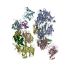
| Title | Structure of active Janus Kinase (JAK) dimer complexed with cytokine receptor intracellular domain | ||||||||||||
 Components Components |
| ||||||||||||
 Keywords Keywords | SIGNALING PROTEIN / signaling complex / Janus Kinase / JAK / oncogenic mutation / gain-of-function mutation / cytokine receptor | ||||||||||||
| Function / homology |  Function and homology information Function and homology informationresponse to type III interferon / interleukin-28 receptor complex / Interleukin-20 family signaling / mucosal immune response / positive regulation of cellular respiration / protein localization to cell-cell junction / interleukin-11-mediated signaling pathway / CCR5 chemokine receptor binding / interleukin-7-mediated signaling pathway / interleukin-9-mediated signaling pathway ...response to type III interferon / interleukin-28 receptor complex / Interleukin-20 family signaling / mucosal immune response / positive regulation of cellular respiration / protein localization to cell-cell junction / interleukin-11-mediated signaling pathway / CCR5 chemokine receptor binding / interleukin-7-mediated signaling pathway / interleukin-9-mediated signaling pathway / interleukin-4-mediated signaling pathway / interleukin-10-mediated signaling pathway / positive regulation of homotypic cell-cell adhesion / regulation of defense response to virus by host / interleukin-15-mediated signaling pathway / growth hormone receptor binding / cytokine receptor activity / extrinsic component of cytoplasmic side of plasma membrane / interleukin-6-mediated signaling pathway / interleukin-2-mediated signaling pathway / type I interferon-mediated signaling pathway / positive regulation of sprouting angiogenesis / type II interferon-mediated signaling pathway / cell surface receptor signaling pathway via JAK-STAT / endomembrane system / non-specific protein-tyrosine kinase / non-membrane spanning protein tyrosine kinase activity / positive regulation of protein localization to nucleus / cytokine-mediated signaling pathway / protein phosphatase binding / defense response to virus / cytoskeleton / cell differentiation / signaling receptor complex / intracellular signal transduction / response to antibiotic / ubiquitin protein ligase binding / regulation of transcription by RNA polymerase II / ATP binding / metal ion binding / nucleus / plasma membrane / cytoplasm Similarity search - Function | ||||||||||||
| Biological species |  | ||||||||||||
| Method | ELECTRON MICROSCOPY / single particle reconstruction / cryo EM / Resolution: 3.6 Å | ||||||||||||
 Authors Authors | Glassman, C.R. / Tsutsumi, N. / Jude, K.M. / Garcia, K.C. | ||||||||||||
| Funding support |  United States, 3items United States, 3items
| ||||||||||||
 Citation Citation | Journal: Science / Year: 2022 Title: Structure of a Janus kinase cytokine receptor complex reveals the basis for dimeric activation. Authors: Caleb R Glassman / Naotaka Tsutsumi / Robert A Saxton / Patrick J Lupardus / Kevin M Jude / K Christopher Garcia / Abstract: Cytokines signal through cell surface receptor dimers to initiate activation of intracellular Janus kinases (JAKs). We report the 3.6-angstrom-resolution cryo-electron microscopy structure of full- ...Cytokines signal through cell surface receptor dimers to initiate activation of intracellular Janus kinases (JAKs). We report the 3.6-angstrom-resolution cryo-electron microscopy structure of full-length JAK1 complexed with a cytokine receptor intracellular domain Box1 and Box2 regions captured as an activated homodimer bearing the valine→phenylalanine (VF) mutation prevalent in myeloproliferative neoplasms. The seven domains of JAK1 form an extended structural unit, the dimerization of which is mediated by close-packing of the pseudokinase (PK) domains from the monomeric subunits. The oncogenic VF mutation lies within the core of the JAK1 PK interdimer interface, enhancing packing complementarity to facilitate ligand-independent activation. The carboxy-terminal tyrosine kinase domains are poised for transactivation and to phosphorylate the receptor STAT (signal transducer and activator of transcription)-recruiting motifs projecting from the overhanging FERM (four-point-one, ezrin, radixin, moesin)-SH2 (Src homology 2)-domains. Mapping of constitutively active JAK mutants supports a two-step allosteric activation mechanism and reveals opportunities for selective therapeutic targeting of oncogenic JAK signaling. | ||||||||||||
| History |
|
- Structure visualization
| Movie |
Movie viewer |
|---|---|
| Structure viewer | Molecule: MolmilJmol/JSmol |
- Downloads & links
-Download
| PDBx/mmCIF format | 7t6f.cif.gz | 394.5 KB | Display | PDBx/mmCIF format |
|---|---|---|---|---|
| PDB format | pdb7t6f.ent.gz | 311.8 KB | Display | PDB format |
| PDBx/mmJSON format | 7t6f.json.gz | Tree view | PDBx/mmJSON format | |
| Others |  Other downloads Other downloads |
-Validation report
| Arichive directory | https://data.pdbj.org/pub/pdb/validation_reports/t6/7t6fftp://data.pdbj.org/pub/pdb/validation_reports/t6/7t6f | HTTPS FTP |
|---|
-Related structure data
| Related structure data |  25715MC M: map data used to model this data C: citing same article ( |
|---|---|
| Similar structure data |


-Links
 PDBj
PDBj


- Assembly
| Deposited unit | 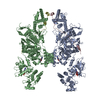
|
|---|---|
| 1 |
|
-Components
| #1: Protein | Mass: 136026.844 Da / Num. of mol.: 2 / Mutation: V657F Source method: isolated from a genetically manipulated source Source: (gene. exp.)  Trichoplusia ni (cabbage looper) Trichoplusia ni (cabbage looper)References: UniProt: B1ASP2, non-specific protein-tyrosine kinase #2: Protein | Mass: 9895.373 Da / Num. of mol.: 2 Source method: isolated from a genetically manipulated source Source: (gene. exp.) Trichoplusia ni (cabbage looper) / References: UniProt: Q8CGK5#3: Chemical |   Mass: 267.241 Da / Num. of mol.: 2 / Source method: obtained synthetically / Formula: C10H13N5O4 Mass: 267.241 Da / Num. of mol.: 2 / Source method: obtained synthetically / Formula: C10H13N5O4#4: Chemical |   Mass: 427.201 Da / Num. of mol.: 2 / Source method: obtained synthetically / Formula: C10H15N5O10P2 / Comment: ADP, energy-carrying molecule*YM Mass: 427.201 Da / Num. of mol.: 2 / Source method: obtained synthetically / Formula: C10H15N5O10P2 / Comment: ADP, energy-carrying molecule*YMHas ligand of interest | N | |
|---|
-Experimental details
-Experiment
| Experiment | Method: ELECTRON MICROSCOPY |
|---|---|
| EM experiment | Aggregation state: PARTICLE / 3D reconstruction method: single particle reconstruction |
- Sample preparation
| Component | Name: GCN4-zippered dimeric IFN-lambda intracellular domain bound to two Jak1s Type: COMPLEX Details: Co-expressed GST-fused GCN4-IFN-lambda and full-length Jak1 in T. ni. GST tagged was removed by 3C protease digestion. Entity ID: #1-#2 / Source: RECOMBINANT | ||||||||||||||||||||||||
|---|---|---|---|---|---|---|---|---|---|---|---|---|---|---|---|---|---|---|---|---|---|---|---|---|---|
| Molecular weight | Value: 0.29 MDa / Experimental value: NO | ||||||||||||||||||||||||
| Source (natural) | Organism: | ||||||||||||||||||||||||
| Source (recombinant) | Organism: Trichoplusia ni (cabbage looper) | ||||||||||||||||||||||||
| Buffer solution | pH: 8 / Details: additive: 0.01% w/v DDM | ||||||||||||||||||||||||
| Buffer component |
| ||||||||||||||||||||||||
| Specimen | Conc.: 6 mg/ml / Embedding applied: NO / Shadowing applied: NO / Staining applied: NO / Vitrification applied: YES / Details: GCN4-mIFN-lambda-box1box2-mJak1 V657F | ||||||||||||||||||||||||
| Specimen support | Grid material: GOLD / Grid mesh size: 300 divisions/in. / Grid type: Quantifoil R1.2/1.3 | ||||||||||||||||||||||||
| Vitrification | Instrument: LEICA EM GP / Cryogen name: ETHANE / Humidity: 95 % / Chamber temperature: 293 K / Details: 3 s blotting before plunging |
- Electron microscopy imaging
| Experimental equipment |  Model: Titan Krios / Image courtesy: FEI Company |
|---|---|
| Microscopy | Model: FEI TITAN KRIOS |
| Electron gun | Electron source:  FIELD EMISSION GUN / Accelerating voltage: 300 kV / Illumination mode: FLOOD BEAM FIELD EMISSION GUN / Accelerating voltage: 300 kV / Illumination mode: FLOOD BEAM |
| Electron lens | Mode: BRIGHT FIELD / Nominal magnification: 29000 X / Calibrated magnification: 58680 X / Nominal defocus max: 2000 nm / Nominal defocus min: 1000 nm / Cs: 2.7 mm / C2 aperture diameter: 100 µm / Alignment procedure: COMA FREE |
| Specimen holder | Cryogen: NITROGEN / Specimen holder model: FEI TITAN KRIOS AUTOGRID HOLDER |
| Image recording | Electron dose: 55 e/Å2 / Detector mode: SUPER-RESOLUTION / Film or detector model: GATAN K3 (6k x 4k) / Num. of grids imaged: 2 / Num. of real images: 29467 |
- Processing
| Software | Name: PHENIX / Version: 1.19.2_4158: / Classification: refinement | ||||||||||||||||||||||||||||||||
|---|---|---|---|---|---|---|---|---|---|---|---|---|---|---|---|---|---|---|---|---|---|---|---|---|---|---|---|---|---|---|---|---|---|
| EM software |
| ||||||||||||||||||||||||||||||||
| CTF correction | Details: Final per-particle CTF values were determined by cryoSPARC Local CTF Refinement. Type: PHASE FLIPPING AND AMPLITUDE CORRECTION | ||||||||||||||||||||||||||||||||
| Particle selection | Num. of particles selected: 17057371 Details: Including non-proteinous features. The actual number of intact complex particles was ~1,429,325. | ||||||||||||||||||||||||||||||||
| Symmetry | Point symmetry: C2 (2 fold cyclic) | ||||||||||||||||||||||||||||||||
| 3D reconstruction | Resolution: 3.6 Å / Resolution method: FSC 0.143 CUT-OFF / Num. of particles: 224615 / Symmetry type: POINT | ||||||||||||||||||||||||||||||||
| Atomic model building | Protocol: FLEXIBLE FIT / Space: REAL | ||||||||||||||||||||||||||||||||
| Refine LS restraints |
|
