 Movie
Movie Controller


+ Open data

- Basic information
| Entry | Database: PDB / ID: 7t60 | ||||||
|---|---|---|---|---|---|---|---|
| Title | P. aeruginosa LpxA in complex with ligand L13 | ||||||
 Components Components | Acyl-[acyl-carrier-protein]--UDP-N-acetylglucosamine O-acyltransferase | ||||||
 Keywords Keywords | TRANSFERASE / Lpxa / lipid A / LPS / lipopolysaccharide / UDP-N-acetylglucosamine O-acyltransferase | ||||||
| Function / homology |  Function and homology information Function and homology informationacyl-[acyl-carrier-protein]-UDP-N-acetylglucosamine O-acyltransferase / acyl-[acyl-carrier-protein]-UDP-N-acetylglucosamine O-acyltransferase activity / lipid A biosynthetic process / membrane / cytoplasm Similarity search - Function | ||||||
| Biological species |  Pseudomonas aeruginosa PA7 (bacteria) Pseudomonas aeruginosa PA7 (bacteria) | ||||||
| Method |  X-RAY DIFFRACTION / SYNCHROTRON / MOLECULAR REPLACEMENT / Resolution: 2 Å X-RAY DIFFRACTION / SYNCHROTRON / MOLECULAR REPLACEMENT / Resolution: 2 Å | ||||||
 Authors Authors | Sacco, M. / Chen, Y. | ||||||
| Funding support | 1items
| ||||||
 Citation Citation | Journal: Acs Infect Dis. / Year: 2022 Title: Structure-Based Ligand Design Targeting Pseudomonas aeruginosa LpxA in Lipid A Biosynthesis. Authors: Sacco, M.D. / Defrees, K. / Zhang, X. / Lawless, W. / Nwanochie, E. / Balsizer, A. / Darch, S.E. / Renslo, A.R. / Chen, Y. | ||||||
| History |
|
- Structure visualization
| Structure viewer | Molecule: MolmilJmol/JSmol |
|---|
- Downloads & links
-Download
| PDBx/mmCIF format | 7t60.cif.gz | 312.5 KB | Display | PDBx/mmCIF format |
|---|---|---|---|---|
| PDB format | pdb7t60.ent.gz | 254 KB | Display | PDB format |
| PDBx/mmJSON format | 7t60.json.gz | Tree view | PDBx/mmJSON format | |
| Others |  Other downloads Other downloads |
-Validation report
| Summary document | 7t60_validation.pdf.gz | 3.3 MB | Display | wwPDB validaton report |
|---|---|---|---|---|
| Full document | 7t60_full_validation.pdf.gz | 3.4 MB | Display | |
| Data in XML | 7t60_validation.xml.gz | 62.1 KB | Display | |
| Data in CIF | 7t60_validation.cif.gz | 82.2 KB | Display | |
| Arichive directory | https://data.pdbj.org/pub/pdb/validation_reports/t6/7t60ftp://data.pdbj.org/pub/pdb/validation_reports/t6/7t60 | HTTPS FTP |
-Related structure data
| Related structure data |  7t5rC  7t5sC  7t5xC  7t5zC  7t61C  6uegS S: Starting model for refinement C: citing same article ( |
|---|---|
| Similar structure data |
-Links
 PDBj
PDBj


- Assembly
| Deposited unit | 
| ||||||||
|---|---|---|---|---|---|---|---|---|---|
| 1 | 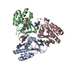
| ||||||||
| 2 | 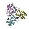
| ||||||||
| Unit cell |
|
-Components
| #1: Protein | Mass: 28048.730 Da / Num. of mol.: 6 Source method: isolated from a genetically manipulated source Source: (gene. exp.) Pseudomonas aeruginosa PA7 (bacteria) / Strain: PA7 / Gene: lpxA, PSPA7_1495Production host: References: UniProt: A6V1E4, acyl-[acyl-carrier-protein]-UDP-N-acetylglucosamine O-acyltransferase #2: Chemical |   Mass: 92.094 Da / Num. of mol.: 2 / Source method: obtained synthetically / Formula: C3H8O3 Mass: 92.094 Da / Num. of mol.: 2 / Source method: obtained synthetically / Formula: C3H8O3#3: Chemical | ChemComp-F9H / (   Mass: 515.445 Da / Num. of mol.: 8 / Source method: obtained synthetically / Formula: C23H20F3N7O4 / Feature type: SUBJECT OF INVESTIGATION Mass: 515.445 Da / Num. of mol.: 8 / Source method: obtained synthetically / Formula: C23H20F3N7O4 / Feature type: SUBJECT OF INVESTIGATION#4: Chemical | ChemComp-TRS / |   Mass: 122.143 Da / Num. of mol.: 1 / Source method: obtained synthetically / Formula: C4H12NO3 / Comment: pH buffer*YM Mass: 122.143 Da / Num. of mol.: 1 / Source method: obtained synthetically / Formula: C4H12NO3 / Comment: pH buffer*YM#5: Water | ChemComp-HOH / |  Mass: 18.015 Da / Num. of mol.: 290 / Source method: isolated from a natural source / Formula: H2O Mass: 18.015 Da / Num. of mol.: 290 / Source method: isolated from a natural source / Formula: H2OHas ligand of interest | Y | |
|---|
-Experimental details
-Experiment
| Experiment | Method: X-RAY DIFFRACTION / Number of used crystals: 1 |
|---|
- Sample preparation
| Crystal | Density Matthews: 2.17 Å3/Da / Density % sol: 43.37 % |
|---|---|
| Crystal grow | Temperature: 293 K / Method: vapor diffusion, hanging drop / Details: 12% PEG 1,000, 0.2 M CaOAc, 0.1 M imidazole pH 7 |
-Data collection
| Diffraction | Mean temperature: 100 K / Serial crystal experiment: N | |||||||||||||||||||||||||||
|---|---|---|---|---|---|---|---|---|---|---|---|---|---|---|---|---|---|---|---|---|---|---|---|---|---|---|---|---|
| Diffraction source | Source: SYNCHROTRON / Site: APS  / Beamline: 19-ID / Wavelength: 0.9789 Å / Beamline: 19-ID / Wavelength: 0.9789 Å | |||||||||||||||||||||||||||
| Detector | Type: DECTRIS PILATUS3 6M / Detector: PIXEL / Date: Dec 10, 2020 | |||||||||||||||||||||||||||
| Radiation | Protocol: SINGLE WAVELENGTH / Monochromatic (M) / Laue (L): M / Scattering type: x-ray | |||||||||||||||||||||||||||
| Radiation wavelength | Wavelength: 0.9789 Å / Relative weight: 1 | |||||||||||||||||||||||||||
| Reflection | Resolution: 2→46.04 Å / Num. obs: 98941 / % possible obs: 99.2 % / Redundancy: 8.3 % / CC1/2: 0.998 / Rmerge(I) obs: 0.082 / Rpim(I) all: 0.03 / Rrim(I) all: 0.088 / Net I/σ(I): 12.6 | |||||||||||||||||||||||||||
| Reflection shell | Diffraction-ID: 1 / % possible all: 97.9
|
- Processing
| Software |
| ||||||||||||||||||||||||||||||||||||||||||||||||||||||||||||
|---|---|---|---|---|---|---|---|---|---|---|---|---|---|---|---|---|---|---|---|---|---|---|---|---|---|---|---|---|---|---|---|---|---|---|---|---|---|---|---|---|---|---|---|---|---|---|---|---|---|---|---|---|---|---|---|---|---|---|---|---|---|
| Refinement | Method to determine structure: MOLECULAR REPLACEMENT Starting model: 6ueg Resolution: 2→46.04 Å / Cor.coef. Fo:Fc: 0.964 / Cor.coef. Fo:Fc free: 0.958 / SU B: 4.673 / SU ML: 0.124 / Cross valid method: THROUGHOUT / σ(F): 0 / ESU R: 0.208 / ESU R Free: 0.162 / Stereochemistry target values: MAXIMUM LIKELIHOOD Details: HYDROGENS HAVE BEEN ADDED IN THE RIDING POSITIONS U VALUES : REFINED INDIVIDUALLY
| ||||||||||||||||||||||||||||||||||||||||||||||||||||||||||||
| Solvent computation | Ion probe radii: 0.8 Å / Shrinkage radii: 0.8 Å / VDW probe radii: 1.2 Å / Solvent model: MASK | ||||||||||||||||||||||||||||||||||||||||||||||||||||||||||||
| Displacement parameters | Biso max: 131.27 Å2 / Biso mean: 42.993 Å2 / Biso min: 18.64 Å2
| ||||||||||||||||||||||||||||||||||||||||||||||||||||||||||||
| Refinement step | Cycle: final / Resolution: 2→46.04 Å
| ||||||||||||||||||||||||||||||||||||||||||||||||||||||||||||
| Refine LS restraints |
| ||||||||||||||||||||||||||||||||||||||||||||||||||||||||||||
| LS refinement shell | Resolution: 2→2.052 Å / Rfactor Rfree error: 0 / Total num. of bins used: 20
|
