 ムービー
ムービー コントローラー


+ データを開く

- 基本情報
基本情報
| 登録情報 | データベース: PDB / ID: 7t5z | ||||||
|---|---|---|---|---|---|---|---|
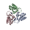
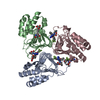
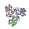
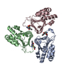
| タイトル | P. aeruginosa LpxA in complex with ligand L8 | ||||||
 要素 要素 | Acyl-[acyl-carrier-protein]--UDP-N-acetylglucosamine O-acyltransferase | ||||||
 キーワード キーワード | TRANSFERASE / Lpxa / lipid A / LPS / lipopolysaccharide / UDP-N-acetylglucosamine O-acyltransferase | ||||||
| 機能・相同性 |  機能・相同性情報 機能・相同性情報acyl-[acyl-carrier-protein]-UDP-N-acetylglucosamine O-acyltransferase / acyl-[acyl-carrier-protein]-UDP-N-acetylglucosamine O-acyltransferase activity / lipid A biosynthetic process / membrane / cytoplasm 類似検索 - 分子機能 | ||||||
| 生物種 |  Pseudomonas aeruginosa PA7 (バクテリア) Pseudomonas aeruginosa PA7 (バクテリア) | ||||||
| 手法 |  X線回折 / シンクロトロン / 分子置換 / 解像度: 2.25 Å X線回折 / シンクロトロン / 分子置換 / 解像度: 2.25 Å | ||||||
 データ登録者 データ登録者 | Sacco, M. / Chen, Y. | ||||||
| 資金援助 | 1件
| ||||||
 引用 引用 | ジャーナル: Acs Infect Dis. / 年: 2022 タイトル: Structure-Based Ligand Design Targeting Pseudomonas aeruginosa LpxA in Lipid A Biosynthesis. 著者: Sacco, M.D. / Defrees, K. / Zhang, X. / Lawless, W. / Nwanochie, E. / Balsizer, A. / Darch, S.E. / Renslo, A.R. / Chen, Y. | ||||||
| 履歴 |
|
- 構造の表示
構造の表示
| 構造ビューア | 分子: MolmilJmol/JSmol |
|---|
- ダウンロードとリンク
-ダウンロード
| PDBx/mmCIF形式 | 7t5z.cif.gz | 309.5 KB | 表示 | PDBx/mmCIF形式 |
|---|---|---|---|---|
| PDB形式 | pdb7t5z.ent.gz | 250.6 KB | 表示 | PDB形式 |
| PDBx/mmJSON形式 | 7t5z.json.gz | ツリー表示 | PDBx/mmJSON形式 | |
| その他 |  その他のダウンロード その他のダウンロード |
-検証レポート
| 文書・要旨 | 7t5z_validation.pdf.gz | 2.5 MB | 表示 | wwPDB検証レポート |
|---|---|---|---|---|
| 文書・詳細版 | 7t5z_full_validation.pdf.gz | 2.5 MB | 表示 | |
| XML形式データ | 7t5z_validation.xml.gz | 59 KB | 表示 | |
| CIF形式データ | 7t5z_validation.cif.gz | 80.8 KB | 表示 | |
| アーカイブディレクトリ | https://data.pdbj.org/pub/pdb/validation_reports/t5/7t5zftp://data.pdbj.org/pub/pdb/validation_reports/t5/7t5z | HTTPS FTP |
-関連構造データ


-リンク
 PDBj
PDBj


- 集合体
集合体
| 登録構造単位 | 
| ||||||||
|---|---|---|---|---|---|---|---|---|---|
| 1 | 
| ||||||||
| 2 | 
| ||||||||
| 単位格子 |
|
-要素
| #1: タンパク質 | 分子量: 28048.730 Da / 分子数: 6 / 由来タイプ: 組換発現 由来: (組換発現) Pseudomonas aeruginosa PA7 (バクテリア)株: PA7 / 遺伝子: lpxA, PSPA7_1495 発現宿主: 参照: UniProt: A6V1E4, acyl-[acyl-carrier-protein]-UDP-N-acetylglucosamine O-acyltransferase #2: 化合物 | ChemComp-F6I / (   分子量: 452.410 Da / 分子数: 6 / 由来タイプ: 合成 / 式: C18H15F3N6O3S / タイプ: SUBJECT OF INVESTIGATION 分子量: 452.410 Da / 分子数: 6 / 由来タイプ: 合成 / 式: C18H15F3N6O3S / タイプ: SUBJECT OF INVESTIGATION#3: 化合物 | ChemComp-PEG / |   分子量: 106.120 Da / 分子数: 1 / 由来タイプ: 合成 / 式: C4H10O3 分子量: 106.120 Da / 分子数: 1 / 由来タイプ: 合成 / 式: C4H10O3#4: 水 | ChemComp-HOH / |  分子量: 18.015 Da / 分子数: 351 / 由来タイプ: 天然 / 式: H2O 分子量: 18.015 Da / 分子数: 351 / 由来タイプ: 天然 / 式: H2O研究の焦点であるリガンドがあるか | Y | |
|---|
-実験情報
-実験
| 実験 | 手法: X線回折 / 使用した結晶の数: 1 |
|---|
- 試料調製
試料調製
| 結晶 | マシュー密度: 2.17 Å3/Da / 溶媒含有率: 43.41 % |
|---|---|
| 結晶化 | 温度: 293 K / 手法: 蒸気拡散法, ハンギングドロップ法 / 詳細: 12% PEG 1,000, 0.2 M CaOAc, 0.1 M imidazole pH 7 |
-データ収集
| 回折 | 平均測定温度: 100 K / Serial crystal experiment: N | ||||||||||||||||||||||||||||||
|---|---|---|---|---|---|---|---|---|---|---|---|---|---|---|---|---|---|---|---|---|---|---|---|---|---|---|---|---|---|---|---|
| 放射光源 | 由来: シンクロトロン / サイト: APS  / ビームライン: 19-BM / 波長: 0.9793 Å / ビームライン: 19-BM / 波長: 0.9793 Å | ||||||||||||||||||||||||||||||
| 検出器 | タイプ: ADSC QUANTUM 210r / 検出器: CCD / 日付: 2019年4月11日 | ||||||||||||||||||||||||||||||
| 放射 | プロトコル: SINGLE WAVELENGTH / 単色(M)・ラウエ(L): M / 散乱光タイプ: x-ray | ||||||||||||||||||||||||||||||
| 放射波長 | 波長: 0.9793 Å / 相対比: 1 | ||||||||||||||||||||||||||||||
| 反射 | 解像度: 2.25→46.04 Å / Num. obs: 56934 / % possible obs: 80.5 % / 冗長度: 4.8 % / CC1/2: 0.996 / Rmerge(I) obs: 0.092 / Rpim(I) all: 0.045 / Rrim(I) all: 0.103 / Net I/σ(I): 11.6 | ||||||||||||||||||||||||||||||
| 反射 シェル | Diffraction-ID: 1
|
- 解析
解析
| ソフトウェア |
| ||||||||||||||||||||||||||||||||||||||||||||||||||||||||||||
|---|---|---|---|---|---|---|---|---|---|---|---|---|---|---|---|---|---|---|---|---|---|---|---|---|---|---|---|---|---|---|---|---|---|---|---|---|---|---|---|---|---|---|---|---|---|---|---|---|---|---|---|---|---|---|---|---|---|---|---|---|---|
| 精密化 | 構造決定の手法: 分子置換 開始モデル: 6ueg 解像度: 2.25→46.04 Å / Cor.coef. Fo:Fc: 0.952 / Cor.coef. Fo:Fc free: 0.935 / SU B: 7.753 / SU ML: 0.184 / 交差検証法: THROUGHOUT / σ(F): 0 / ESU R: 0.756 / ESU R Free: 0.267 / 立体化学のターゲット値: MAXIMUM LIKELIHOOD 詳細: HYDROGENS HAVE BEEN ADDED IN THE RIDING POSITIONS U VALUES : REFINED INDIVIDUALLY
| ||||||||||||||||||||||||||||||||||||||||||||||||||||||||||||
| 溶媒の処理 | イオンプローブ半径: 0.8 Å / 減衰半径: 0.8 Å / VDWプローブ半径: 1.2 Å / 溶媒モデル: MASK | ||||||||||||||||||||||||||||||||||||||||||||||||||||||||||||
| 原子変位パラメータ | Biso max: 123.24 Å2 / Biso mean: 36.274 Å2 / Biso min: 12.51 Å2
| ||||||||||||||||||||||||||||||||||||||||||||||||||||||||||||
| 精密化ステップ | サイクル: final / 解像度: 2.25→46.04 Å
| ||||||||||||||||||||||||||||||||||||||||||||||||||||||||||||
| 拘束条件 |
| ||||||||||||||||||||||||||||||||||||||||||||||||||||||||||||
| LS精密化 シェル | 解像度: 2.25→2.308 Å / Rfactor Rfree error: 0 / Total num. of bins used: 20
|
