 ムービー
ムービー コントローラー


+ データを開く

- 基本情報
基本情報
| 登録情報 | データベース: PDB / ID: 7t3s | ||||||
|---|---|---|---|---|---|---|---|
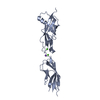
| タイトル | Crystal Structure of Mouse Cadherin-23 EC8-9 | ||||||
 要素 要素 | Cadherin-23 | ||||||
 キーワード キーワード | CELL ADHESION / HEARING / MECHANOTRANSDUCTION / CALCIUM-BINDING PROTEIN | ||||||
| 機能・相同性 |  機能・相同性情報 機能・相同性情報equilibrioception / sensory perception of light stimulus / cochlear hair cell ribbon synapse / stereocilium tip / inner ear receptor cell stereocilium organization / photoreceptor ribbon synapse / kinocilium / inner ear auditory receptor cell differentiation / calcium-dependent cell-cell adhesion / stereocilium ...equilibrioception / sensory perception of light stimulus / cochlear hair cell ribbon synapse / stereocilium tip / inner ear receptor cell stereocilium organization / photoreceptor ribbon synapse / kinocilium / inner ear auditory receptor cell differentiation / calcium-dependent cell-cell adhesion / stereocilium / photoreceptor cell maintenance / auditory receptor cell stereocilium organization / inner ear morphogenesis / homophilic cell-cell adhesion / inner ear development / cochlea development / regulation of cytosolic calcium ion concentration / photoreceptor inner segment / locomotory behavior / sensory perception of sound / calcium ion transport / cell adhesion / calcium ion binding / synapse / centrosome / plasma membrane 類似検索 - 分子機能 | ||||||
| 生物種 |  | ||||||
| 手法 |  X線回折 / シンクロトロン / 分子置換 / 解像度: 2.966 Å X線回折 / シンクロトロン / 分子置換 / 解像度: 2.966 Å | ||||||
 データ登録者 データ登録者 | Sandhu, J.S. / De La Torre, P. / Sotomayor, M. | ||||||
| 資金援助 |  米国, 1件 米国, 1件
| ||||||
 引用 引用 | ジャーナル: to be published タイトル: Crystal Structure of Mouse Cadherin-23 EC8-9 著者: Sandhu, J.S. / De La Torre, P. / Sotomayor, M. | ||||||
| 履歴 |
|
- 構造の表示
構造の表示
| 構造ビューア | 分子: MolmilJmol/JSmol |
|---|
- ダウンロードとリンク
-ダウンロード
| PDBx/mmCIF形式 | 7t3s.cif.gz | 59.6 KB | 表示 | PDBx/mmCIF形式 |
|---|---|---|---|---|
| PDB形式 | pdb7t3s.ent.gz | 38.4 KB | 表示 | PDB形式 |
| PDBx/mmJSON形式 | 7t3s.json.gz | ツリー表示 | PDBx/mmJSON形式 | |
| その他 |  その他のダウンロード その他のダウンロード |
-検証レポート
| 文書・要旨 | 7t3s_validation.pdf.gz | 1.2 MB | 表示 | wwPDB検証レポート |
|---|---|---|---|---|
| 文書・詳細版 | 7t3s_full_validation.pdf.gz | 1.2 MB | 表示 | |
| XML形式データ | 7t3s_validation.xml.gz | 9.2 KB | 表示 | |
| CIF形式データ | 7t3s_validation.cif.gz | 11.6 KB | 表示 | |
| アーカイブディレクトリ | https://data.pdbj.org/pub/pdb/validation_reports/t3/7t3sftp://data.pdbj.org/pub/pdb/validation_reports/t3/7t3s | HTTPS FTP |
-関連構造データ

-リンク
 PDBj
PDBj
- 集合体
集合体
| 登録構造単位 | 
| ||||||||
|---|---|---|---|---|---|---|---|---|---|
| 1 |
| ||||||||
| 単位格子 |
|
-要素
| #1: タンパク質 | 分子量: 25550.609 Da / 分子数: 1 / 由来タイプ: 組換発現 / 由来: (組換発現)  | ||||||
|---|---|---|---|---|---|---|---|
| #2: 化合物 |   分子量: 40.078 Da / 分子数: 3 / 由来タイプ: 合成 / 式: Ca / タイプ: SUBJECT OF INVESTIGATION 分子量: 40.078 Da / 分子数: 3 / 由来タイプ: 合成 / 式: Ca / タイプ: SUBJECT OF INVESTIGATION#3: 化合物 | ChemComp-NA / |   分子量: 22.990 Da / 分子数: 1 / 由来タイプ: 合成 / 式: Na 分子量: 22.990 Da / 分子数: 1 / 由来タイプ: 合成 / 式: Na#4: 水 | ChemComp-HOH / |  分子量: 18.015 Da / 分子数: 1 / 由来タイプ: 天然 / 式: H2O 分子量: 18.015 Da / 分子数: 1 / 由来タイプ: 天然 / 式: H2O研究の焦点であるリガンドがあるか | Y | |
-実験情報
-実験
| 実験 | 手法: X線回折 / 使用した結晶の数: 1 |
|---|
- 試料調製
試料調製
| 結晶 | マシュー密度: 2.16 Å3/Da / 溶媒含有率: 43.07 % |
|---|---|
| 結晶化 | 温度: 277 K / 手法: 蒸気拡散法, シッティングドロップ法 / 詳細: 0.2 M NaCl 20% PEG 3350 |
-データ収集
| 回折 | 平均測定温度: 100 K / Serial crystal experiment: N |
|---|---|
| 放射光源 | 由来: シンクロトロン / サイト: APS / ビームライン: 24-ID-E / 波長: 0.97918 Å |
| 検出器 | タイプ: DECTRIS EIGER X 16M / 検出器: PIXEL / 日付: 2018年11月8日 |
| 放射 | プロトコル: SINGLE WAVELENGTH / 単色(M)・ラウエ(L): M / 散乱光タイプ: x-ray |
| 放射波長 | 波長: 0.97918 Å / 相対比: 1 |
| 反射 | 解像度: 2.966→48.213 Å / Num. obs: 4947 / % possible obs: 98.9 % / 冗長度: 8.8 % / CC1/2: 0.966 / Net I/σ(I): 7.75 |
| 反射 シェル | 解像度: 2.97→3.02 Å / 冗長度: 4.5 % / Mean I/σ(I) obs: 1.77 / Num. unique obs: 224 / CC1/2: 0.809 / % possible all: 95.3 |
- 解析
解析
| ソフトウェア |
| |||||||||||||||||||||||||||||||||||||||||||||||||||||||||||||||||||||||||||||||||||||||||||||||||||||||||||||||||||||||||||||||||||||||||||||||||||||||||||||||||||||||||||||||||||||||||||||||||||||||||||||||||||||||||||||||||||||||
|---|---|---|---|---|---|---|---|---|---|---|---|---|---|---|---|---|---|---|---|---|---|---|---|---|---|---|---|---|---|---|---|---|---|---|---|---|---|---|---|---|---|---|---|---|---|---|---|---|---|---|---|---|---|---|---|---|---|---|---|---|---|---|---|---|---|---|---|---|---|---|---|---|---|---|---|---|---|---|---|---|---|---|---|---|---|---|---|---|---|---|---|---|---|---|---|---|---|---|---|---|---|---|---|---|---|---|---|---|---|---|---|---|---|---|---|---|---|---|---|---|---|---|---|---|---|---|---|---|---|---|---|---|---|---|---|---|---|---|---|---|---|---|---|---|---|---|---|---|---|---|---|---|---|---|---|---|---|---|---|---|---|---|---|---|---|---|---|---|---|---|---|---|---|---|---|---|---|---|---|---|---|---|---|---|---|---|---|---|---|---|---|---|---|---|---|---|---|---|---|---|---|---|---|---|---|---|---|---|---|---|---|---|---|---|---|---|---|---|---|---|---|---|---|---|---|---|---|---|---|---|---|---|
| 精密化 | 構造決定の手法: 分子置換 開始モデル: 5tfl,5tfk 解像度: 2.966→48.213 Å / Cor.coef. Fo:Fc: 0.923 / Cor.coef. Fo:Fc free: 0.832 / SU B: 24.59 / SU ML: 0.446 / 交差検証法: FREE R-VALUE / ESU R Free: 0.538 / 詳細: Hydrogens have been added in their riding positions
| |||||||||||||||||||||||||||||||||||||||||||||||||||||||||||||||||||||||||||||||||||||||||||||||||||||||||||||||||||||||||||||||||||||||||||||||||||||||||||||||||||||||||||||||||||||||||||||||||||||||||||||||||||||||||||||||||||||||
| 溶媒の処理 | イオンプローブ半径: 0.8 Å / 減衰半径: 0.8 Å / VDWプローブ半径: 1.2 Å / 溶媒モデル: MASK BULK SOLVENT | |||||||||||||||||||||||||||||||||||||||||||||||||||||||||||||||||||||||||||||||||||||||||||||||||||||||||||||||||||||||||||||||||||||||||||||||||||||||||||||||||||||||||||||||||||||||||||||||||||||||||||||||||||||||||||||||||||||||
| 原子変位パラメータ | Biso mean: 52.1 Å2
| |||||||||||||||||||||||||||||||||||||||||||||||||||||||||||||||||||||||||||||||||||||||||||||||||||||||||||||||||||||||||||||||||||||||||||||||||||||||||||||||||||||||||||||||||||||||||||||||||||||||||||||||||||||||||||||||||||||||
| 精密化ステップ | サイクル: LAST / 解像度: 2.966→48.213 Å
| |||||||||||||||||||||||||||||||||||||||||||||||||||||||||||||||||||||||||||||||||||||||||||||||||||||||||||||||||||||||||||||||||||||||||||||||||||||||||||||||||||||||||||||||||||||||||||||||||||||||||||||||||||||||||||||||||||||||
| 拘束条件 |
| |||||||||||||||||||||||||||||||||||||||||||||||||||||||||||||||||||||||||||||||||||||||||||||||||||||||||||||||||||||||||||||||||||||||||||||||||||||||||||||||||||||||||||||||||||||||||||||||||||||||||||||||||||||||||||||||||||||||
| LS精密化 シェル | Refine-ID: X-RAY DIFFRACTION / Total num. of bins used: 20
|
