 Movie
Movie Controller


+ Open data

- Basic information
| Entry | Database: PDB / ID: 7t3i | ||||||
|---|---|---|---|---|---|---|---|
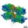
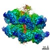
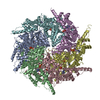
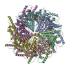
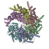
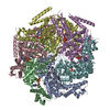
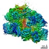
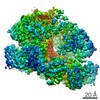
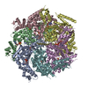
| Title | CryoEM structure of the Rix7 D2 Walker B mutant | ||||||
 Components Components |
| ||||||
 Keywords Keywords | RIBOSOMAL PROTEIN / AAA-ATPase / Rix7 / substrate translocation | ||||||
| Function / homology |  Function and homology information Function and homology informationpreribosome binding / peroxisome organization / ribosome biogenesis / ATP hydrolysis activity / RNA binding / ATP binding / nucleus Similarity search - Function | ||||||
| Biological species |  Chaetomium thermophilum (fungus) Chaetomium thermophilum (fungus) | ||||||
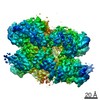
| Method | ELECTRON MICROSCOPY / single particle reconstruction / cryo EM / Resolution: 4.3 Å | ||||||
 Authors Authors | Kocaman, S. / Stanley, R.E. | ||||||
| Funding support |  United States, 1items United States, 1items
| ||||||
 Citation Citation | Journal: PNAS Nexus / Year: 2022 Title: Communication network within the essential AAA-ATPase Rix7 drives ribosome assembly. Authors: Seda Kocaman / Yu-Hua Lo / Juno M Krahn / Mack Sobhany / Venkata P Dandey / Matthew L Petrovich / Suhas K Etigunta / Jason G Williams / Leesa J Deterding / Mario J Borgnia / Robin E Stanley / Abstract: Rix7 is an essential AAA+ ATPase that functions during the early stages of ribosome biogenesis. Rix7 is composed of three domains including an N-terminal domain (NTD) and two AAA+ domains (D1 and ...Rix7 is an essential AAA+ ATPase that functions during the early stages of ribosome biogenesis. Rix7 is composed of three domains including an N-terminal domain (NTD) and two AAA+ domains (D1 and D2) that assemble into an asymmetric stacked hexamer. It was recently established that Rix7 is a presumed protein translocase that removes substrates from preribosomes by translocating them through its central pore. However, how the different domains of Rix7 coordinate their activities within the overall hexameric structure was unknown. We captured cryo-electron microscopy (EM) structures of single and double Walker B variants of full length Rix7. The disordered NTD was not visible in the cryo-EM reconstructions, but cross-linking mass spectrometry revealed that the NTD can associate with the central channel in vitro. Deletion of the disordered NTD enabled us to obtain a structure of the Rix7 hexamer to 2.9 Å resolution, providing high resolution details of critical motifs involved in substrate translocation and interdomain communication. This structure coupled with cell-based assays established that the linker connecting the D1 and D2 domains as well as the pore loops lining the central channel are essential for formation of the large ribosomal subunit. Together, our work shows that Rix7 utilizes a complex communication network to drive ribosome biogenesis. | ||||||
| History |
|
- Structure visualization
| Movie |
Movie viewer |
|---|---|
| Structure viewer | Molecule: MolmilJmol/JSmol |
- Downloads & links
-Download
| PDBx/mmCIF format | 7t3i.cif.gz | 608.3 KB | Display | PDBx/mmCIF format |
|---|---|---|---|---|
| PDB format | pdb7t3i.ent.gz | 486 KB | Display | PDB format |
| PDBx/mmJSON format | 7t3i.json.gz | Tree view | PDBx/mmJSON format | |
| Others |  Other downloads Other downloads |
-Validation report
| Arichive directory | https://data.pdbj.org/pub/pdb/validation_reports/t3/7t3iftp://data.pdbj.org/pub/pdb/validation_reports/t3/7t3i | HTTPS FTP |
|---|
-Related structure data
| Related structure data | 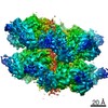 25659MC 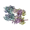 7swlC  7t0vC M: map data used to model this data C: citing same article ( |
|---|---|
| Similar structure data |
-Links
 PDBj
PDBj


- Assembly
| Deposited unit | 
|
|---|---|
| 1 |
|
-Components
| #1: Protein | Mass: 89419.250 Da / Num. of mol.: 6 Source method: isolated from a genetically manipulated source Source: (gene. exp.) Chaetomium thermophilum (fungus) / Gene: CTHT_0002840 / Production host: #2: Protein/peptide | | Mass: 2315.846 Da / Num. of mol.: 1 / Source method: isolated from a natural source / Source: (natural) #3: Chemical | ChemComp-ATP /   Mass: 507.181 Da / Num. of mol.: 8 / Source method: obtained synthetically / Formula: C10H16N5O13P3 / Feature type: SUBJECT OF INVESTIGATION / Comment: ATP, energy-carrying molecule*YM Mass: 507.181 Da / Num. of mol.: 8 / Source method: obtained synthetically / Formula: C10H16N5O13P3 / Feature type: SUBJECT OF INVESTIGATION / Comment: ATP, energy-carrying molecule*YM#4: Chemical | ChemComp-PO4 / |   Mass: 94.971 Da / Num. of mol.: 1 / Source method: obtained synthetically / Formula: PO4 / Feature type: SUBJECT OF INVESTIGATION Mass: 94.971 Da / Num. of mol.: 1 / Source method: obtained synthetically / Formula: PO4 / Feature type: SUBJECT OF INVESTIGATIONHas ligand of interest | Y | |
|---|
-Experimental details
-Experiment
| Experiment | Method: ELECTRON MICROSCOPY |
|---|---|
| EM experiment | Aggregation state: PARTICLE / 3D reconstruction method: single particle reconstruction |
- Sample preparation
| Component | Name: CryoEM structure of Rix7 D2 Walker B mutant / Type: COMPLEX / Entity ID: #1-#2 / Source: RECOMBINANT |
|---|---|
| Source (natural) | Organism: Chaetomium thermophilum (fungus) |
| Source (recombinant) | Organism: |
| Buffer solution | pH: 8 |
| Specimen | Embedding applied: NO / Shadowing applied: NO / Staining applied: NO / Vitrification applied: YES |
| Vitrification | Cryogen name: ETHANE |
- Electron microscopy imaging
| Experimental equipment |  Model: Titan Krios / Image courtesy: FEI Company |
|---|---|
| Microscopy | Model: FEI TITAN KRIOS |
| Electron gun | Electron source:  FIELD EMISSION GUN / Accelerating voltage: 300 kV / Illumination mode: FLOOD BEAM FIELD EMISSION GUN / Accelerating voltage: 300 kV / Illumination mode: FLOOD BEAM |
| Electron lens | Mode: BRIGHT FIELD / Nominal defocus max: 2700 nm / Nominal defocus min: 1200 nm |
| Image recording | Electron dose: 40 e/Å2 / Film or detector model: GATAN K3 (6k x 4k) |
- Processing
| Software |
| ||||||||||||||||||||||||
|---|---|---|---|---|---|---|---|---|---|---|---|---|---|---|---|---|---|---|---|---|---|---|---|---|---|
| CTF correction | Type: PHASE FLIPPING AND AMPLITUDE CORRECTION | ||||||||||||||||||||||||
| 3D reconstruction | Resolution: 4.3 Å / Resolution method: FSC 0.143 CUT-OFF / Num. of particles: 584000 / Symmetry type: POINT | ||||||||||||||||||||||||
| Refinement | Cross valid method: NONE Stereochemistry target values: GeoStd + Monomer Library + CDL v1.2 | ||||||||||||||||||||||||
| Displacement parameters | Biso mean: 90.92 Å2 | ||||||||||||||||||||||||
| Refine LS restraints |
|
