 Movie
Movie Controller

[English] 日本語
 Yorodumi
- PDB-7t27: Structure of phage FBB1 anti-CBASS nuclease Acb1-3'3'-cGAMP compl... -
+ Open data

- Basic information
| Entry | Database: PDB / ID: 7t27 | ||||||
|---|---|---|---|---|---|---|---|
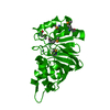
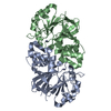
| Title | Structure of phage FBB1 anti-CBASS nuclease Acb1-3'3'-cGAMP complex in post reaction state | ||||||
 Components Components | Acb1 | ||||||
 Keywords Keywords | VIRAL PROTEIN / Anti-CBASS / Nuclease / Immune evasion | ||||||
| Function / homology | Cyclic phosphodiesterase / Chem-ECI / Anti-CBASS protein Acb1 Function and homology information Function and homology information | ||||||
| Biological species |  Erwinia phage FBB1 (virus) Erwinia phage FBB1 (virus) | ||||||
| Method |  X-RAY DIFFRACTION / SYNCHROTRON / SAD / Resolution: 1.2 Å X-RAY DIFFRACTION / SYNCHROTRON / SAD / Resolution: 1.2 Å | ||||||
 Authors Authors | Hobbs, S.J. / Wein, T. / Lu, A. / Morehouse, B.R. / Schnabel, J. / Sorek, R. / Kranzusch, P.J. | ||||||
| Funding support |  United States, 1items United States, 1items
| ||||||
 Citation Citation | Journal: Nature / Year: 2022 Title: Phage anti-CBASS and anti-Pycsar nucleases subvert bacterial immunity. Authors: Hobbs, S.J. / Wein, T. / Lu, A. / Morehouse, B.R. / Schnabel, J. / Leavitt, A. / Yirmiya, E. / Sorek, R. / Kranzusch, P.J. | ||||||
| History |
|
- Structure visualization
| Structure viewer | Molecule: MolmilJmol/JSmol |
|---|
- Downloads & links
-Download
| PDBx/mmCIF format | 7t27.cif.gz | 122.1 KB | Display | PDBx/mmCIF format |
|---|---|---|---|---|
| PDB format | pdb7t27.ent.gz | 79.1 KB | Display | PDB format |
| PDBx/mmJSON format | 7t27.json.gz | Tree view | PDBx/mmJSON format | |
| Others |  Other downloads Other downloads |
-Validation report
| Summary document | 7t27_validation.pdf.gz | 770 KB | Display | wwPDB validaton report |
|---|---|---|---|---|
| Full document | 7t27_full_validation.pdf.gz | 773.6 KB | Display | |
| Data in XML | 7t27_validation.xml.gz | 11.3 KB | Display | |
| Data in CIF | 7t27_validation.cif.gz | 16.3 KB | Display | |
| Arichive directory | https://data.pdbj.org/pub/pdb/validation_reports/t2/7t27ftp://data.pdbj.org/pub/pdb/validation_reports/t2/7t27 | HTTPS FTP |
-Related structure data


-Links
 PDBj
PDBj- Assembly
| Deposited unit | 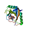
| ||||||||||||
|---|---|---|---|---|---|---|---|---|---|---|---|---|---|
| 1 |
| ||||||||||||
| Unit cell |
|
-Components
| #1: Protein | Mass: 16265.133 Da / Num. of mol.: 1 Source method: isolated from a genetically manipulated source Source: (gene. exp.) Erwinia phage FBB1 (virus) / Production host:  |
|---|---|
| #2: Chemical | ChemComp-ECI / [(  Mass: 724.558 Da / Num. of mol.: 1 / Source method: obtained synthetically / Formula: C20H26N10O12P2S2 / Feature type: SUBJECT OF INVESTIGATION Mass: 724.558 Da / Num. of mol.: 1 / Source method: obtained synthetically / Formula: C20H26N10O12P2S2 / Feature type: SUBJECT OF INVESTIGATION |
| #3: Chemical | ChemComp-SO4 /   Mass: 96.063 Da / Num. of mol.: 1 / Source method: obtained synthetically / Formula: SO4 Mass: 96.063 Da / Num. of mol.: 1 / Source method: obtained synthetically / Formula: SO4 |
| #4: Water | ChemComp-HOH /  Mass: 18.015 Da / Num. of mol.: 220 / Source method: isolated from a natural source / Formula: H2O Mass: 18.015 Da / Num. of mol.: 220 / Source method: isolated from a natural source / Formula: H2O |
| Has ligand of interest | Y |
-Experimental details
-Experiment
| Experiment | Method: X-RAY DIFFRACTION / Number of used crystals: 1 |
|---|
- Sample preparation
| Crystal | Density Matthews: 2.28 Å3/Da / Density % sol: 45.99 % |
|---|---|
| Crystal grow | Temperature: 291 K / Method: vapor diffusion, hanging drop / Details: 2 M ammonium sulfate, 0.1 M sodium citrate pH 4.6 |
-Data collection
| Diffraction | Mean temperature: 80 K / Serial crystal experiment: N |
|---|---|
| Diffraction source | Source: SYNCHROTRON / Site: APS / Beamline: 24-ID-C / Wavelength: 0.97918 Å |
| Detector | Type: DECTRIS EIGER2 X 16M / Detector: PIXEL / Date: Sep 24, 2021 |
| Radiation | Protocol: SINGLE WAVELENGTH / Monochromatic (M) / Laue (L): M / Scattering type: x-ray |
| Radiation wavelength | Wavelength: 0.97918 Å / Relative weight: 1 |
| Reflection | Resolution: 1.2→43.85 Å / Num. obs: 47842 / % possible obs: 99.1 % / Redundancy: 16.9 % / Biso Wilson estimate: 9.31 Å2 / CC1/2: 1 / Rpim(I) all: 0.013 / Net I/σ(I): 23.2 |
| Reflection shell | Resolution: 1.2→1.22 Å / Mean I/σ(I) obs: 1.4 / Num. unique obs: 2307 / CC1/2: 0.846 / Rpim(I) all: 0.438 / % possible all: 94.9 |
- Processing
| Software |
| |||||||||||||||||||||||||||||||||||||||||||||||||||||||||||||||||||||||||||||||||||||||||||||||||||||||||||||||||||||||||||||||||||||||||||||||||||||||||||||||||||||||||||||||
|---|---|---|---|---|---|---|---|---|---|---|---|---|---|---|---|---|---|---|---|---|---|---|---|---|---|---|---|---|---|---|---|---|---|---|---|---|---|---|---|---|---|---|---|---|---|---|---|---|---|---|---|---|---|---|---|---|---|---|---|---|---|---|---|---|---|---|---|---|---|---|---|---|---|---|---|---|---|---|---|---|---|---|---|---|---|---|---|---|---|---|---|---|---|---|---|---|---|---|---|---|---|---|---|---|---|---|---|---|---|---|---|---|---|---|---|---|---|---|---|---|---|---|---|---|---|---|---|---|---|---|---|---|---|---|---|---|---|---|---|---|---|---|---|---|---|---|---|---|---|---|---|---|---|---|---|---|---|---|---|---|---|---|---|---|---|---|---|---|---|---|---|---|---|---|---|---|
| Refinement | Method to determine structure: SAD / Resolution: 1.2→42.17 Å / SU ML: 0.1195 / Cross valid method: FREE R-VALUE / σ(F): 1.36 / Phase error: 18.2934 Stereochemistry target values: GeoStd + Monomer Library + CDL v1.2
| |||||||||||||||||||||||||||||||||||||||||||||||||||||||||||||||||||||||||||||||||||||||||||||||||||||||||||||||||||||||||||||||||||||||||||||||||||||||||||||||||||||||||||||||
| Solvent computation | Shrinkage radii: 0.9 Å / VDW probe radii: 1.11 Å / Solvent model: FLAT BULK SOLVENT MODEL | |||||||||||||||||||||||||||||||||||||||||||||||||||||||||||||||||||||||||||||||||||||||||||||||||||||||||||||||||||||||||||||||||||||||||||||||||||||||||||||||||||||||||||||||
| Displacement parameters | Biso mean: 14.6 Å2 | |||||||||||||||||||||||||||||||||||||||||||||||||||||||||||||||||||||||||||||||||||||||||||||||||||||||||||||||||||||||||||||||||||||||||||||||||||||||||||||||||||||||||||||||
| Refinement step | Cycle: LAST / Resolution: 1.2→42.17 Å
| |||||||||||||||||||||||||||||||||||||||||||||||||||||||||||||||||||||||||||||||||||||||||||||||||||||||||||||||||||||||||||||||||||||||||||||||||||||||||||||||||||||||||||||||
| Refine LS restraints |
| |||||||||||||||||||||||||||||||||||||||||||||||||||||||||||||||||||||||||||||||||||||||||||||||||||||||||||||||||||||||||||||||||||||||||||||||||||||||||||||||||||||||||||||||
| LS refinement shell |
| |||||||||||||||||||||||||||||||||||||||||||||||||||||||||||||||||||||||||||||||||||||||||||||||||||||||||||||||||||||||||||||||||||||||||||||||||||||||||||||||||||||||||||||||
| Refinement TLS params. | Method: refined / Refine-ID: X-RAY DIFFRACTION
| |||||||||||||||||||||||||||||||||||||||||||||||||||||||||||||||||||||||||||||||||||||||||||||||||||||||||||||||||||||||||||||||||||||||||||||||||||||||||||||||||||||||||||||||
| Refinement TLS group | Refine-ID: X-RAY DIFFRACTION / Auth asym-ID: A / Label asym-ID: A
|
