Movie
Movie Controller


+ Open data

- Basic information
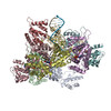
| Entry | Database: PDB / ID: 7svv | ||||||
|---|---|---|---|---|---|---|---|
| Title | TnsBctd-TnsC complex | ||||||
 Components Components |
| ||||||
 Keywords Keywords | DNA BINDING PROTEIN/DNA / CAST / transposase / AAA+ ATPase / AAA+ / CRISPR / Cas / DNA BINDING PROTEIN-DNA complex | ||||||
| Function / homology | PHOSPHOAMINOPHOSPHONIC ACID-ADENYLATE ESTER / DNA / DNA (> 10) Function and homology information Function and homology information | ||||||
| Biological species |  [Scytonema hofmanni] UTEX 2349 (bacteria) [Scytonema hofmanni] UTEX 2349 (bacteria)synthetic construct (others) | ||||||
| Method | ELECTRON MICROSCOPY / helical reconstruction / cryo EM / Resolution: 3.54 Å | ||||||
 Authors Authors | Park, J. / Tsai, A.W.T. / Kellogg, E.H. | ||||||
| Funding support |  United States, 1items United States, 1items
| ||||||
 Citation Citation | Journal: Proc Natl Acad Sci U S A / Year: 2022 Title: Mechanistic details of CRISPR-associated transposon recruitment and integration revealed by cryo-EM. Authors: Jung-Un Park / Amy Wei-Lun Tsai / Tiffany H Chen / Joseph E Peters / Elizabeth H Kellogg / Abstract: CRISPR-associated transposons (CASTs) are Tn7-like elements that are capable of RNA-guided DNA integration. Although structural data are known for nearly all core transposition components, the ...CRISPR-associated transposons (CASTs) are Tn7-like elements that are capable of RNA-guided DNA integration. Although structural data are known for nearly all core transposition components, the transposase component, TnsB, remains uncharacterized. Using cryo-electron microscopy (cryo-EM) structure determination, we reveal the conformation of TnsB during transposon integration for the type V-K CAST system from (ShCAST). Our structure of TnsB is a tetramer, revealing strong mechanistic relationships with the overall architecture of RNaseH transposases/integrases in general, and in particular the MuA transposase from bacteriophage Mu. However, key structural differences in the C-terminal domains indicate that TnsB's tetrameric architecture is stabilized by a different set of protein-protein interactions compared with MuA. We describe the base-specific interactions along the TnsB binding site, which explain how different CAST elements can function on cognate mobile elements independent of one another. We observe that melting of the 5' nontransferred strand of the transposon end is a structural feature stabilized by TnsB and furthermore is crucial for donor-DNA integration. Although not observed in the TnsB strand-transfer complex, the C-terminal end of TnsB serves a crucial role in transposase recruitment to the target site. The C-terminal end of TnsB adopts a short, structured 15-residue "hook" that decorates TnsC filaments. Unlike full-length TnsB, C-terminal fragments do not appear to stimulate filament disassembly using two different assays, suggesting that additional interactions between TnsB and TnsC are required for redistributing TnsC to appropriate targets. The structural information presented here will help guide future work in modifying these important systems as programmable gene integration tools. | ||||||
| History |
|
- Structure visualization
| Structure viewer | Molecule: MolmilJmol/JSmol |
|---|
- Downloads & links
-Download
| PDBx/mmCIF format | 7svv.cif.gz | 486.3 KB | Display | PDBx/mmCIF format |
|---|---|---|---|---|
| PDB format | pdb7svv.ent.gz | 389.9 KB | Display | PDB format |
| PDBx/mmJSON format | 7svv.json.gz | Tree view | PDBx/mmJSON format | |
| Others |  Other downloads Other downloads |
-Validation report
| Arichive directory | https://data.pdbj.org/pub/pdb/validation_reports/sv/7svvftp://data.pdbj.org/pub/pdb/validation_reports/sv/7svv | HTTPS FTP |
|---|
-Related structure data
| Related structure data |  25454MC 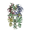 7svwC M: map data used to model this data C: citing same article ( |
|---|---|
| Similar structure data |


-Links
 PDBj
PDBj


- Assembly
| Deposited unit | 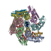
|
|---|---|
| 1 |
|
-Components
-DNA chain , 2 types, 2 molecules 12
| #1: DNA chain | Mass: 5734.706 Da / Num. of mol.: 1 / Source method: obtained synthetically / Source: (synth.) synthetic construct (others) |
|---|---|
| #2: DNA chain | Mass: 6845.593 Da / Num. of mol.: 1 / Source method: obtained synthetically / Source: (synth.) synthetic construct (others) |
-Protein / Protein/peptide , 2 types, 20 molecules ABCDEFGHIJabcdefghij
| #3: Protein | Mass: 31444.617 Da / Num. of mol.: 10 Source method: isolated from a genetically manipulated source Source: (gene. exp.) [Scytonema hofmanni] UTEX 2349 (bacteria)Production host: #4: Protein/peptide | Mass: 1977.110 Da / Num. of mol.: 10 Source method: isolated from a genetically manipulated source Source: (gene. exp.) [Scytonema hofmanni] UTEX 2349 (bacteria)Production host: |
|---|
-Non-polymers , 2 types, 20 molecules 

| #5: Chemical | ChemComp-ANP /  Mass: 506.196 Da / Num. of mol.: 10 / Source method: obtained synthetically / Formula: C10H17N6O12P3 / Comment: AMP-PNP, energy-carrying molecule analogue*YM Mass: 506.196 Da / Num. of mol.: 10 / Source method: obtained synthetically / Formula: C10H17N6O12P3 / Comment: AMP-PNP, energy-carrying molecule analogue*YM#6: Chemical | ChemComp-MG /  Mass: 24.305 Da / Num. of mol.: 10 / Source method: obtained synthetically / Formula: Mg Mass: 24.305 Da / Num. of mol.: 10 / Source method: obtained synthetically / Formula: Mg |
|---|
-Details
| Has ligand of interest | N |
|---|
-Experimental details
-Experiment
| Experiment | Method: ELECTRON MICROSCOPY |
|---|---|
| EM experiment | Aggregation state: PARTICLE / 3D reconstruction method: helical reconstruction |
- Sample preparation
| Component | Name: AMPPNP-bound TnsBctd-TnsC filament from ShCAST element Type: COMPLEX / Entity ID: #1-#4 / Source: MULTIPLE SOURCES | |||||||||||||||||||||||||||||||||||
|---|---|---|---|---|---|---|---|---|---|---|---|---|---|---|---|---|---|---|---|---|---|---|---|---|---|---|---|---|---|---|---|---|---|---|---|---|
| Molecular weight | Value: 0.4 MDa / Experimental value: NO | |||||||||||||||||||||||||||||||||||
| Source (natural) | Organism: [Scytonema hofmanni] UTEX 2349 (bacteria) | |||||||||||||||||||||||||||||||||||
| Source (recombinant) | Organism: | |||||||||||||||||||||||||||||||||||
| Buffer solution | pH: 7.5 | |||||||||||||||||||||||||||||||||||
| Buffer component |
| |||||||||||||||||||||||||||||||||||
| Specimen | Conc.: 1 mg/ml / Embedding applied: NO / Shadowing applied: NO / Staining applied: NO / Vitrification applied: YES | |||||||||||||||||||||||||||||||||||
| Specimen support | Grid type: UltrAuFoil R1.2/1.3 | |||||||||||||||||||||||||||||||||||
| Vitrification | Instrument: FEI VITROBOT MARK IV / Cryogen name: ETHANE / Humidity: 100 % / Chamber temperature: 277.15 K |
- Electron microscopy imaging
| Experimental equipment |  Model: Talos Arctica / Image courtesy: FEI Company |
|---|---|
| Microscopy | Model: FEI TALOS ARCTICA |
| Electron gun | Electron source:  FIELD EMISSION GUN / Accelerating voltage: 200 kV / Illumination mode: FLOOD BEAM FIELD EMISSION GUN / Accelerating voltage: 200 kV / Illumination mode: FLOOD BEAM |
| Electron lens | Mode: BRIGHT FIELD / Nominal defocus max: 2500 nm / Nominal defocus min: 1000 nm |
| Specimen holder | Cryogen: NITROGEN / Specimen holder model: FEI TITAN KRIOS AUTOGRID HOLDER |
| Image recording | Electron dose: 50 e/Å2 / Film or detector model: GATAN K3 BIOQUANTUM (6k x 4k) |
- Processing
| EM software |
| |||||||||||||||
|---|---|---|---|---|---|---|---|---|---|---|---|---|---|---|---|---|
| CTF correction | Type: PHASE FLIPPING AND AMPLITUDE CORRECTION | |||||||||||||||
| Helical symmerty | Angular rotation/subunit: 59.3 ° / Axial rise/subunit: 7 Å / Axial symmetry: C1 | |||||||||||||||
| 3D reconstruction | Resolution: 3.54 Å / Resolution method: FSC 0.143 CUT-OFF / Num. of particles: 286988 / Symmetry type: HELICAL | |||||||||||||||
| Atomic model building | Protocol: FLEXIBLE FIT | |||||||||||||||
| Atomic model building | PDB-ID: 7M99 Pdb chain-ID: A / Accession code: 7M99 / Source name: PDB / Type: experimental model |