 Movie
Movie Controller

[English] 日本語
 Yorodumi
- PDB-7sqn: Structure of the E. coli PutA proline dehydrogenase domain (resid... -
+ Open data

- Basic information
| Entry | Database: PDB / ID: 7sqn | ||||||
|---|---|---|---|---|---|---|---|
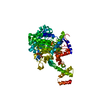
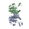
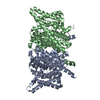
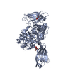
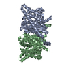
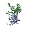
| Title | Structure of the E. coli PutA proline dehydrogenase domain (residues 86-630) complexed with (2S)-oxetane-2-carboxylic acid | ||||||
 Components Components | Bifunctional protein PutA | ||||||
 Keywords Keywords | OXIDOREDUCTASE / BETA/ALPHA BARREL / FLAVOENZYME / PROLINE CATABOLISM | ||||||
| Function / homology | (2S)-oxetane-2-carboxylic acid / FLAVIN-ADENINE DINUCLEOTIDE / TRIETHYLENE GLYCOL / :  Function and homology information Function and homology information | ||||||
| Biological species |  | ||||||
| Method |  X-RAY DIFFRACTION / SYNCHROTRON / FOURIER SYNTHESIS / Resolution: 2.25 Å X-RAY DIFFRACTION / SYNCHROTRON / FOURIER SYNTHESIS / Resolution: 2.25 Å | ||||||
 Authors Authors | Tanner, J.J. / Bogner, A.N. | ||||||
| Funding support |  United States, 1items United States, 1items
| ||||||
 Citation Citation | Journal: Org.Biomol.Chem. / Year: 2022 Title: Structure-affinity relationships of reversible proline analog inhibitors targeting proline dehydrogenase. Authors: Bogner, A.N. / Tanner, J.J. | ||||||
| History |
|
- Structure visualization
| Structure viewer | Molecule: MolmilJmol/JSmol |
|---|
- Downloads & links
-Download
| PDBx/mmCIF format | 7sqn.cif.gz | 210 KB | Display | PDBx/mmCIF format |
|---|---|---|---|---|
| PDB format | pdb7sqn.ent.gz | 163.8 KB | Display | PDB format |
| PDBx/mmJSON format | 7sqn.json.gz | Tree view | PDBx/mmJSON format | |
| Others |  Other downloads Other downloads |
-Validation report
| Arichive directory | https://data.pdbj.org/pub/pdb/validation_reports/sq/7sqnftp://data.pdbj.org/pub/pdb/validation_reports/sq/7sqn | HTTPS FTP |
|---|
-Related structure data
| Related structure data |  7mwtC 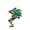 7mwuSC  7mwvC S: Starting model for refinement C: citing same article ( |
|---|---|
| Similar structure data |


-Links
 PDBj
PDBj- Assembly
| Deposited unit | 
| ||||||||
|---|---|---|---|---|---|---|---|---|---|
| 1 |
| ||||||||
| Unit cell |
| ||||||||
| Components on special symmetry positions |
|
-Components
-Protein , 1 types, 1 molecules A
| #1: Protein | Mass: 61451.102 Da / Num. of mol.: 1 / Fragment: residues 86-630 Source method: isolated from a genetically manipulated source Source: (gene. exp.) References: UniProt: A0A383H020, proline dehydrogenase, L-glutamate gamma-semialdehyde dehydrogenase |
|---|
-Non-polymers , 5 types, 62 molecules 


| #2: Chemical | ChemComp-FAD /  Mass: 785.550 Da / Num. of mol.: 1 / Source method: obtained synthetically / Formula: C27H33N9O15P2 / Feature type: SUBJECT OF INVESTIGATION / Comment: FAD*YM Mass: 785.550 Da / Num. of mol.: 1 / Source method: obtained synthetically / Formula: C27H33N9O15P2 / Feature type: SUBJECT OF INVESTIGATION / Comment: FAD*YM |
|---|---|
| #3: Chemical | ChemComp-PGE /  Mass: 150.173 Da / Num. of mol.: 1 / Source method: obtained synthetically / Formula: C6H14O4 Mass: 150.173 Da / Num. of mol.: 1 / Source method: obtained synthetically / Formula: C6H14O4 |
| #4: Chemical | ChemComp-1PE /  Mass: 238.278 Da / Num. of mol.: 1 / Source method: obtained synthetically / Formula: C10H22O6 / Comment: precipitant*YM Mass: 238.278 Da / Num. of mol.: 1 / Source method: obtained synthetically / Formula: C10H22O6 / Comment: precipitant*YM |
| #5: Chemical | ChemComp-A8G / ( Mass: 102.089 Da / Num. of mol.: 1 / Source method: obtained synthetically / Formula: C4H6O3 / Feature type: SUBJECT OF INVESTIGATION Mass: 102.089 Da / Num. of mol.: 1 / Source method: obtained synthetically / Formula: C4H6O3 / Feature type: SUBJECT OF INVESTIGATION |
| #6: Water | ChemComp-HOH / Mass: 18.015 Da / Num. of mol.: 58 / Source method: isolated from a natural source / Formula: H2O |
-Details
| Has ligand of interest | Y |
|---|
-Experimental details
-Experiment
| Experiment | Method: X-RAY DIFFRACTION / Number of used crystals: 1 |
|---|
- Sample preparation
| Crystal | Density Matthews: 2.81 Å3/Da / Density % sol: 56.24 % |
|---|---|
| Crystal grow | Temperature: 295 K / Method: vapor diffusion, hanging drop Details: 100 mM sodium citrate pH 5.6, 5.75, 5.9 or 6.0 and 18-28% PEG 3000 |
-Data collection
| Diffraction | Mean temperature: 100 K / Serial crystal experiment: N | ||||||||||||||||||||||||||||||
|---|---|---|---|---|---|---|---|---|---|---|---|---|---|---|---|---|---|---|---|---|---|---|---|---|---|---|---|---|---|---|---|
| Diffraction source | Source: SYNCHROTRON / Site: APS / Beamline: 24-ID-E / Wavelength: 0.97918 Å | ||||||||||||||||||||||||||||||
| Detector | Type: DECTRIS EIGER X 16M / Detector: PIXEL / Date: Jul 3, 2021 | ||||||||||||||||||||||||||||||
| Radiation | Protocol: SINGLE WAVELENGTH / Monochromatic (M) / Laue (L): M / Scattering type: x-ray | ||||||||||||||||||||||||||||||
| Radiation wavelength | Wavelength: 0.97918 Å / Relative weight: 1 | ||||||||||||||||||||||||||||||
| Reflection | Resolution: 2.25→64.72 Å / Num. obs: 35985 / % possible obs: 99.8 % / Redundancy: 5.3 % / Biso Wilson estimate: 55.05 Å2 / CC1/2: 0.993 / Rmerge(I) obs: 0.114 / Rpim(I) all: 0.053 / Rrim(I) all: 0.126 / Net I/σ(I): 6.3 / Num. measured all: 191098 / Scaling rejects: 86 | ||||||||||||||||||||||||||||||
| Reflection shell | Diffraction-ID: 1
|
- Processing
| Software |
| ||||||||||||||||||||||||||||||||||||||||||||||||||||||||||||||||||||||||||||||||||||||||||||||||||
|---|---|---|---|---|---|---|---|---|---|---|---|---|---|---|---|---|---|---|---|---|---|---|---|---|---|---|---|---|---|---|---|---|---|---|---|---|---|---|---|---|---|---|---|---|---|---|---|---|---|---|---|---|---|---|---|---|---|---|---|---|---|---|---|---|---|---|---|---|---|---|---|---|---|---|---|---|---|---|---|---|---|---|---|---|---|---|---|---|---|---|---|---|---|---|---|---|---|---|---|
| Refinement | Method to determine structure: FOURIER SYNTHESIS Starting model: 7MWU Resolution: 2.25→64.72 Å / SU ML: 0.36 / Cross valid method: THROUGHOUT / σ(F): 1.35 / Phase error: 29.62 / Stereochemistry target values: ML
| ||||||||||||||||||||||||||||||||||||||||||||||||||||||||||||||||||||||||||||||||||||||||||||||||||
| Solvent computation | Shrinkage radii: 0.9 Å / VDW probe radii: 1.11 Å / Solvent model: FLAT BULK SOLVENT MODEL | ||||||||||||||||||||||||||||||||||||||||||||||||||||||||||||||||||||||||||||||||||||||||||||||||||
| Displacement parameters | Biso max: 153.66 Å2 / Biso mean: 69.6152 Å2 / Biso min: 37.42 Å2 | ||||||||||||||||||||||||||||||||||||||||||||||||||||||||||||||||||||||||||||||||||||||||||||||||||
| Refinement step | Cycle: final / Resolution: 2.25→64.72 Å
| ||||||||||||||||||||||||||||||||||||||||||||||||||||||||||||||||||||||||||||||||||||||||||||||||||
| LS refinement shell | Refine-ID: X-RAY DIFFRACTION / Rfactor Rfree error: 0 / Total num. of bins used: 13
| ||||||||||||||||||||||||||||||||||||||||||||||||||||||||||||||||||||||||||||||||||||||||||||||||||
| Refinement TLS params. | Method: refined / Origin x: 4.9795 Å / Origin y: 49.4039 Å / Origin z: 49.3375 Å
| ||||||||||||||||||||||||||||||||||||||||||||||||||||||||||||||||||||||||||||||||||||||||||||||||||
| Refinement TLS group | Selection details: chain A and peptide |
