 Movie
Movie Controller

[English] 日本語
 Yorodumi
- PDB-7sqi: Crosslinked Crystal Structure of Type II Fatty Acid Synthase Keto... -
+ Open data

- Basic information
| Entry | Database: PDB / ID: 7sqi | ||||||
|---|---|---|---|---|---|---|---|
| Title | Crosslinked Crystal Structure of Type II Fatty Acid Synthase Ketosynthase, FabB, and C14-crypto Acyl Carrier Protein, AcpP | ||||||
 Components Components |
| ||||||
 Keywords Keywords | TRANSFERASE / ketosynthase / crosslink / FabB / ACP / KASI | ||||||
| Function / homology |  Function and homology information Function and homology informationlipid A biosynthetic process / acyl binding / acyl carrier activity / membrane / cytosol Similarity search - Function | ||||||
| Biological species |  | ||||||
| Method |  X-RAY DIFFRACTION / SYNCHROTRON / MOLECULAR REPLACEMENT / Resolution: 1.7 Å X-RAY DIFFRACTION / SYNCHROTRON / MOLECULAR REPLACEMENT / Resolution: 1.7 Å | ||||||
 Authors Authors | Chen, A. / Mindrebo, J.T. / Davis, T.D. / Noel, J.P. / Burkart, M.D. | ||||||
| Funding support |  United States, 1items United States, 1items
| ||||||
 Citation Citation | Journal: Acta Crystallogr D Struct Biol / Year: 2022 Title: Mechanism-based cross-linking probes capture the Escherichia coli ketosynthase FabB in conformationally distinct catalytic states. Authors: Chen, A. / Mindrebo, J.T. / Davis, T.D. / Kim, W.E. / Katsuyama, Y. / Jiang, Z. / Ohnishi, Y. / Noel, J.P. / Burkart, M.D. | ||||||
| History |
|
- Structure visualization
| Structure viewer | Molecule: MolmilJmol/JSmol |
|---|
- Downloads & links
-Download
| PDBx/mmCIF format | 7sqi.cif.gz | 223.8 KB | Display | PDBx/mmCIF format |
|---|---|---|---|---|
| PDB format | pdb7sqi.ent.gz | 174.5 KB | Display | PDB format |
| PDBx/mmJSON format | 7sqi.json.gz | Tree view | PDBx/mmJSON format | |
| Others |  Other downloads Other downloads |
-Validation report
| Arichive directory | https://data.pdbj.org/pub/pdb/validation_reports/sq/7sqiftp://data.pdbj.org/pub/pdb/validation_reports/sq/7sqi | HTTPS FTP |
|---|
-Related structure data
| Related structure data |  7sz9C  6okcS S: Starting model for refinement C: citing same article ( |
|---|---|
| Similar structure data |
-Links
 PDBj
PDBj
- Assembly
| Deposited unit | 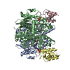
| ||||||||
|---|---|---|---|---|---|---|---|---|---|
| 1 |
| ||||||||
| Unit cell |
|
-Components
| #1: Protein | Mass: 42541.117 Da / Num. of mol.: 2 Source method: isolated from a genetically manipulated source Source: (gene. exp.) References: UniProt: A0A6D2VX38, beta-ketoacyl-[acyl-carrier-protein] synthase I #2: Protein | Mass: 8645.460 Da / Num. of mol.: 2 Source method: isolated from a genetically manipulated source Source: (gene. exp.) #3: Chemical |   Mass: 22.990 Da / Num. of mol.: 2 / Source method: obtained synthetically / Formula: Na Mass: 22.990 Da / Num. of mol.: 2 / Source method: obtained synthetically / Formula: Na#4: Chemical |   Mass: 584.083 Da / Num. of mol.: 2 / Source method: obtained synthetically / Formula: C25H47ClN3O8P Mass: 584.083 Da / Num. of mol.: 2 / Source method: obtained synthetically / Formula: C25H47ClN3O8P#5: Water | ChemComp-HOH / |  Mass: 18.015 Da / Num. of mol.: 1069 / Source method: isolated from a natural source / Formula: H2O Mass: 18.015 Da / Num. of mol.: 1069 / Source method: isolated from a natural source / Formula: H2OHas ligand of interest | N | Has protein modification | Y | |
|---|
-Experimental details
-Experiment
| Experiment | Method: X-RAY DIFFRACTION / Number of used crystals: 1 |
|---|
- Sample preparation
| Crystal | Density Matthews: 2.29 Å3/Da / Density % sol: 46.36 % |
|---|---|
| Crystal grow | Temperature: 281 K / Method: vapor diffusion, hanging drop / pH: 6.5 Details: 20% PEG 8000, 0.3 M sodium acetate, 0.1 M sodium cacodylate |
-Data collection
| Diffraction | Mean temperature: 100 K / Serial crystal experiment: N | ||||||||||||||||||||||||
|---|---|---|---|---|---|---|---|---|---|---|---|---|---|---|---|---|---|---|---|---|---|---|---|---|---|
| Diffraction source | Source: SYNCHROTRON / Site: ALS / Beamline: 8.2.1 / Wavelength: 1 Å | ||||||||||||||||||||||||
| Detector | Type: ADSC QUANTUM 315r / Detector: CCD / Date: Jul 3, 2019 | ||||||||||||||||||||||||
| Radiation | Protocol: SINGLE WAVELENGTH / Monochromatic (M) / Laue (L): M / Scattering type: x-ray | ||||||||||||||||||||||||
| Radiation wavelength | Wavelength: 1 Å / Relative weight: 1 | ||||||||||||||||||||||||
| Reflection | Resolution: 1.7→39.1 Å / Num. obs: 104223 / % possible obs: 99.68 % / Redundancy: 12 % / Biso Wilson estimate: 16.6 Å2 / Rmerge(I) obs: 0.342 / Rpim(I) all: 0.102 / Rrim(I) all: 0.357 / Net I/σ(I): 527 | ||||||||||||||||||||||||
| Reflection shell | Diffraction-ID: 1
|
- Processing
| Software |
| ||||||||||||||||||
|---|---|---|---|---|---|---|---|---|---|---|---|---|---|---|---|---|---|---|---|
| Refinement | Method to determine structure: MOLECULAR REPLACEMENT Starting model: 6okc Resolution: 1.7→39.1 Å / Cross valid method: THROUGHOUT
| ||||||||||||||||||
| Displacement parameters | Biso max: 120.72 Å2 / Biso mean: 23.8032 Å2 / Biso min: 7.79 Å2 | ||||||||||||||||||
| Refinement step | Cycle: LAST / Resolution: 1.7→39.1 Å
| ||||||||||||||||||
| LS refinement shell | Resolution: 1.7→1.761 Å /
|
