 Movie
Movie Controller


+ Open data

- Basic information
| Entry | Database: PDB / ID: 7sp3 | ||||||
|---|---|---|---|---|---|---|---|
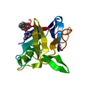
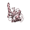
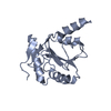
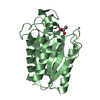
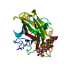
| Title | E. coli RppH bound to Ap4A | ||||||
 Components Components | RNA pyrophosphohydrolase | ||||||
 Keywords Keywords | RNA BINDING PROTEIN / Cap / Ap4A / RppH / Nudix hydrolase | ||||||
| Function / homology |  Function and homology information Function and homology informationmRNA 5'-diphosphatase activity / mRNA catabolic process / Hydrolases; Acting on acid anhydrides; In phosphorus-containing anhydrides / cytoplasm Similarity search - Function | ||||||
| Biological species |  | ||||||
| Method |  X-RAY DIFFRACTION / MOLECULAR REPLACEMENT / Resolution: 1.6 Å X-RAY DIFFRACTION / MOLECULAR REPLACEMENT / Resolution: 1.6 Å | ||||||
 Authors Authors | Serganov, A.A. / Vasilyev, N. / Nuthanakanti, A. | ||||||
| Funding support |  United States, 1items United States, 1items
| ||||||
 Citation Citation | Journal: Proc.Natl.Acad.Sci.USA / Year: 2022 Title: A distinct RNA recognition mechanism governs Np 4 decapping by RppH. Authors: Levenson-Palmer, R. / Luciano, D.J. / Vasilyev, N. / Nuthanakanti, A. / Serganov, A. / Belasco, J.G. | ||||||
| History |
|
- Structure visualization
| Structure viewer | Molecule: MolmilJmol/JSmol |
|---|
- Downloads & links
-Download
| PDBx/mmCIF format | 7sp3.cif.gz | 62.8 KB | Display | PDBx/mmCIF format |
|---|---|---|---|---|
| PDB format | pdb7sp3.ent.gz | 35 KB | Display | PDB format |
| PDBx/mmJSON format | 7sp3.json.gz | Tree view | PDBx/mmJSON format | |
| Others |  Other downloads Other downloads |
-Validation report
| Arichive directory | https://data.pdbj.org/pub/pdb/validation_reports/sp/7sp3ftp://data.pdbj.org/pub/pdb/validation_reports/sp/7sp3 | HTTPS FTP |
|---|
-Related structure data
| Related structure data | 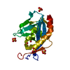 4s2wS S: Starting model for refinement |
|---|---|
| Similar structure data |


-Links
 PDBj
PDBj

- Assembly
| Deposited unit | 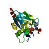
| ||||||||||||
|---|---|---|---|---|---|---|---|---|---|---|---|---|---|
| 1 |
| ||||||||||||
| Unit cell |
|
-Components
-Protein , 1 types, 1 molecules A
| #1: Protein | Mass: 18965.773 Da / Num. of mol.: 1 Source method: isolated from a genetically manipulated source Source: (gene. exp.) Gene: rppH, nudH, D9D94_13650, D9E88_05455, D9J61_06705, G5V60_04980, GP711_07030 Production host: References: UniProt: A0A3K0QF38, Hydrolases; Acting on acid anhydrides; In phosphorus-containing anhydrides |
|---|
-Non-polymers , 5 types, 118 molecules 


| #2: Chemical | ChemComp-B4P /  Mass: 836.387 Da / Num. of mol.: 1 / Source method: obtained synthetically / Formula: C20H28N10O19P4 / Feature type: SUBJECT OF INVESTIGATION Mass: 836.387 Da / Num. of mol.: 1 / Source method: obtained synthetically / Formula: C20H28N10O19P4 / Feature type: SUBJECT OF INVESTIGATION | ||||||
|---|---|---|---|---|---|---|---|
| #3: Chemical |  Mass: 24.305 Da / Num. of mol.: 3 / Source method: obtained synthetically / Formula: Mg Mass: 24.305 Da / Num. of mol.: 3 / Source method: obtained synthetically / Formula: Mg#4: Chemical | ChemComp-F / |  Mass: 18.998 Da / Num. of mol.: 1 / Source method: obtained synthetically / Formula: F Mass: 18.998 Da / Num. of mol.: 1 / Source method: obtained synthetically / Formula: F#5: Chemical | ChemComp-CL / |  Mass: 35.453 Da / Num. of mol.: 1 / Source method: obtained synthetically / Formula: Cl Mass: 35.453 Da / Num. of mol.: 1 / Source method: obtained synthetically / Formula: Cl#6: Water | ChemComp-HOH / | Mass: 18.015 Da / Num. of mol.: 112 / Source method: isolated from a natural source / Formula: H2O |
-Details
| Has ligand of interest | Y |
|---|
-Experimental details
-Experiment
| Experiment | Method: X-RAY DIFFRACTION / Number of used crystals: 1 |
|---|
- Sample preparation
| Crystal | Density Matthews: 2.15 Å3/Da / Density % sol: 42.84 % |
|---|---|
| Crystal grow | Temperature: 293 K / Method: vapor diffusion, hanging drop / pH: 5 Details: 0.4 M (NH4)2SO4, 10% (v/v) PEG3350, and 10% glycerol |
-Data collection
| Diffraction | Mean temperature: 100 K / Serial crystal experiment: N |
|---|---|
| Diffraction source | Source: ROTATING ANODE / Type: RIGAKU R-AXIS / Wavelength: 1.54 Å |
| Detector | Type: RIGAKU / Detector: IMAGE PLATE / Date: Sep 23, 2013 |
| Radiation | Protocol: SINGLE WAVELENGTH / Monochromatic (M) / Laue (L): M / Scattering type: x-ray |
| Radiation wavelength | Wavelength: 1.54 Å / Relative weight: 1 |
| Reflection | Resolution: 1.6→19.4 Å / Num. obs: 20993 / % possible obs: 97.5 % / Redundancy: 5.2 % / Biso Wilson estimate: 18.88 Å2 / CC1/2: 1 / CC star: 1 / Rmerge(I) obs: 0.043 / Rpim(I) all: 0.021 / Rrim(I) all: 0.048 / Net I/σ(I): 22.49 |
| Reflection shell | Resolution: 1.6→1.657 Å / Redundancy: 4.4 % / Rmerge(I) obs: 0.5874 / Mean I/σ(I) obs: 2.6 / Num. unique obs: 1890 / CC1/2: 0.752 / CC star: 0.926 / Rpim(I) all: 0.3048 / Rrim(I) all: 0.6652 / % possible all: 89.32 |
- Processing
| Software |
| ||||||||||||||||||||||||||||||||||||||||||||||||||||||||
|---|---|---|---|---|---|---|---|---|---|---|---|---|---|---|---|---|---|---|---|---|---|---|---|---|---|---|---|---|---|---|---|---|---|---|---|---|---|---|---|---|---|---|---|---|---|---|---|---|---|---|---|---|---|---|---|---|---|
| Refinement | Method to determine structure: MOLECULAR REPLACEMENT Starting model: 4S2W Resolution: 1.6→18.84 Å / SU ML: 0.197 / Cross valid method: FREE R-VALUE / σ(F): 1.37 / Phase error: 24.9407 Stereochemistry target values: GeoStd + Monomer Library + CDL v1.2
| ||||||||||||||||||||||||||||||||||||||||||||||||||||||||
| Solvent computation | Shrinkage radii: 0.9 Å / VDW probe radii: 1.11 Å / Solvent model: FLAT BULK SOLVENT MODEL | ||||||||||||||||||||||||||||||||||||||||||||||||||||||||
| Displacement parameters | Biso mean: 25.77 Å2 | ||||||||||||||||||||||||||||||||||||||||||||||||||||||||
| Refinement step | Cycle: LAST / Resolution: 1.6→18.84 Å
| ||||||||||||||||||||||||||||||||||||||||||||||||||||||||
| Refine LS restraints |
| ||||||||||||||||||||||||||||||||||||||||||||||||||||||||
| LS refinement shell |
|
