 Movie
Movie Controller


+ Open data

- Basic information
| Entry | Database: PDB / ID: 7soy | ||||||||||||
|---|---|---|---|---|---|---|---|---|---|---|---|---|---|
| Title | The structure of the PP2A-B56gamma1 holoenzyme-PME-1 complex | ||||||||||||
 Components Components |
| ||||||||||||
 Keywords Keywords | RECOMBINATION / The structure of PP2A-B56gamma holoenzyme-PME-1 complex | ||||||||||||
| Function / homology |  Function and homology information Function and homology informationprotein phosphatase methylesterase-1 / protein methylesterase activity / meiotic spindle elongation / PP2A-mediated dephosphorylation of key metabolic factors / RNA polymerase II CTD heptapeptide repeat S2 phosphatase activity / RNA polymerase II CTD heptapeptide repeat S7 phosphatase activity / peptidyl-threonine dephosphorylation / regulation of meiotic cell cycle process involved in oocyte maturation / mitotic sister chromatid separation / MASTL Facilitates Mitotic Progression ...protein phosphatase methylesterase-1 / protein methylesterase activity / meiotic spindle elongation / PP2A-mediated dephosphorylation of key metabolic factors / RNA polymerase II CTD heptapeptide repeat S2 phosphatase activity / RNA polymerase II CTD heptapeptide repeat S7 phosphatase activity / peptidyl-threonine dephosphorylation / regulation of meiotic cell cycle process involved in oocyte maturation / mitotic sister chromatid separation / MASTL Facilitates Mitotic Progression / protein phosphatase type 2A complex / meiotic sister chromatid cohesion, centromeric / INTAC complex / RNA polymerase II CTD heptapeptide repeat S5 phosphatase activity / FAR/SIN/STRIPAK complex / female meiotic nuclear division / Regulation of glycolysis by fructose 2,6-bisphosphate metabolism / meiotic sister chromatid cohesion / Inhibition of replication initiation of damaged DNA by RB1/E2F1 / protein phosphatase regulator activity / protein antigen binding / GABA receptor binding / APC truncation mutants have impaired AXIN binding / AXIN missense mutants destabilize the destruction complex / Truncations of AMER1 destabilize the destruction complex / positive regulation of extrinsic apoptotic signaling pathway in absence of ligand / ERKs are inactivated / Initiation of Nuclear Envelope (NE) Reformation / Beta-catenin phosphorylation cascade / Signaling by GSK3beta mutants / CTNNB1 S33 mutants aren't phosphorylated / CTNNB1 S37 mutants aren't phosphorylated / CTNNB1 S45 mutants aren't phosphorylated / CTNNB1 T41 mutants aren't phosphorylated / Co-stimulation by CD28 / RNA polymerase II transcription initiation surveillance / regulation of growth / Disassembly of the destruction complex and recruitment of AXIN to the membrane / protein dephosphorylation / negative regulation of epithelial to mesenchymal transition / Co-inhibition by CTLA4 / Platelet sensitization by LDL / protein phosphatase inhibitor activity / protein-serine/threonine phosphatase / negative regulation of glycolytic process through fructose-6-phosphate / ERK/MAPK targets / lncRNA binding / T cell homeostasis / vascular endothelial cell response to oscillatory fluid shear stress / mesoderm development / protein serine/threonine phosphatase activity / positive regulation of NLRP3 inflammasome complex assembly / regulation of cell differentiation / regulation of microtubule polymerization / lateral plasma membrane / protein phosphatase activator activity / intrinsic apoptotic signaling pathway in response to DNA damage by p53 class mediator / chromosome, centromeric region / DARPP-32 events / negative regulation of hippo signaling / Cyclin A/B1/B2 associated events during G2/M transition / regulation of G1/S transition of mitotic cell cycle / Nonsense Mediated Decay (NMD) enhanced by the Exon Junction Complex (EJC) / spindle assembly / phosphoprotein phosphatase activity / Loss of Nlp from mitotic centrosomes / Loss of proteins required for interphase microtubule organization from the centrosome / Amplification of signal from unattached kinetochores via a MAD2 inhibitory signal / Recruitment of mitotic centrosome proteins and complexes / protein tyrosine phosphatase activity / Recruitment of NuMA to mitotic centrosomes / Anchoring of the basal body to the plasma membrane / Mitotic Prometaphase / EML4 and NUDC in mitotic spindle formation / protein phosphatase 2A binding / Turbulent (oscillatory, disturbed) flow shear stress activates signaling by PIEZO1 and integrins in endothelial cells / AURKA Activation by TPX2 / Resolution of Sister Chromatid Cohesion / negative regulation of phosphatidylinositol 3-kinase/protein kinase B signal transduction / DNA damage response, signal transduction by p53 class mediator / chromosome segregation / negative regulation of canonical Wnt signaling pathway / RAF activation / RHO GTPases Activate Formins / meiotic cell cycle / Spry regulation of FGF signaling / PKR-mediated signaling / response to lead ion / G2/M transition of mitotic cell cycle / tau protein binding / Degradation of beta-catenin by the destruction complex / spindle pole / Cyclin D associated events in G1 / Negative regulation of MAPK pathway / Separation of Sister Chromatids / Regulation of TP53 Degradation / Regulation of PLK1 Activity at G2/M Transition / mitotic cell cycle / microtubule cytoskeleton / PI5P, PP2A and IER3 Regulate PI3K/AKT Signaling Similarity search - Function | ||||||||||||
| Biological species |  Homo sapiens (human) Homo sapiens (human) | ||||||||||||
| Method | ELECTRON MICROSCOPY / single particle reconstruction / cryo EM / Resolution: 3.4 Å | ||||||||||||
 Authors Authors | Li, Y. / Balakrishnan, V.K. / Rowse, M. / Novikova, I.V. / Xing, Y. | ||||||||||||
| Funding support | 3items
| ||||||||||||
 Citation Citation | Journal: Elife / Year: 2022 Title: Coupling to short linear motifs creates versatile PME-1 activities in PP2A holoenzyme demethylation and inhibition. Authors: Yitong Li / Vijaya Kumar Balakrishnan / Michael Rowse / Cheng-Guo Wu / Anastasia Phoebe Bravos / Vikash K Yadav / Ylva Ivarsson / Stefan Strack / Irina V Novikova / Yongna Xing /   Abstract: Protein phosphatase 2A (PP2A) holoenzymes target broad substrates by recognizing short motifs via regulatory subunits. PP2A methylesterase 1 (PME-1) is a cancer-promoting enzyme and undergoes ...Protein phosphatase 2A (PP2A) holoenzymes target broad substrates by recognizing short motifs via regulatory subunits. PP2A methylesterase 1 (PME-1) is a cancer-promoting enzyme and undergoes methylesterase activation upon binding to the PP2A core enzyme. Here, we showed that PME-1 readily demethylates different families of PP2A holoenzymes and blocks substrate recognition in vitro. The high-resolution cryoelectron microscopy structure of a PP2A-B56 holoenzyme-PME-1 complex reveals that PME-1 disordered regions, including a substrate-mimicking motif, tether to the B56 regulatory subunit at remote sites. They occupy the holoenzyme substrate-binding groove and allow large structural shifts in both holoenzyme and PME-1 to enable multipartite contacts at structured cores to activate the methylesterase. B56 interface mutations selectively block PME-1 activity toward PP2A-B56 holoenzymes and affect the methylation of a fraction of total cellular PP2A. The B56 interface mutations allow us to uncover B56-specific PME-1 functions in p53 signaling. Our studies reveal multiple mechanisms of PME-1 in suppressing holoenzyme functions and versatile PME-1 activities derived from coupling substrate-mimicking motifs to dynamic structured cores. #1: Journal: Acta Crystallogr., Sect. D: Biol. Crystallogr. / Year: 2018Title: Real-space refinement in PHENIX for cryo-EM and crystallography Authors: Xing, Y. | ||||||||||||
| History |
|
- Structure visualization
| Structure viewer | Molecule: MolmilJmol/JSmol |
|---|
- Downloads & links
-Download
| PDBx/mmCIF format | 7soy.cif.gz | 281.5 KB | Display | PDBx/mmCIF format |
|---|---|---|---|---|
| PDB format | pdb7soy.ent.gz | 222.4 KB | Display | PDB format |
| PDBx/mmJSON format | 7soy.json.gz | Tree view | PDBx/mmJSON format | |
| Others |  Other downloads Other downloads |
-Validation report
| Arichive directory | https://data.pdbj.org/pub/pdb/validation_reports/so/7soyftp://data.pdbj.org/pub/pdb/validation_reports/so/7soy | HTTPS FTP |
|---|
-Related structure data
| Related structure data |  25363MC M: map data used to model this data C: citing same article ( |
|---|---|
| Similar structure data |
-Links
 PDBj
PDBj


- Assembly
| Deposited unit | 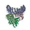
|
|---|---|
| 1 |
|
-Components
| #1: Protein | Mass: 65378.344 Da / Num. of mol.: 1 Source method: isolated from a genetically manipulated source Source: (gene. exp.) Homo sapiens (human) / Gene: PPP2R1A / Production host:  |
|---|---|
| #2: Protein | Mass: 52695.270 Da / Num. of mol.: 1 Source method: isolated from a genetically manipulated source Source: (gene. exp.) Homo sapiens (human) / Gene: PPP2R5C, KIAA0044 / Production host: |
| #3: Protein | Mass: 35636.152 Da / Num. of mol.: 1 Source method: isolated from a genetically manipulated source Source: (gene. exp.) Homo sapiens (human) / Gene: PPP2CA / Production host:  Trichoplusia ni (cabbage looper) Trichoplusia ni (cabbage looper)References: UniProt: P67775, protein-serine/threonine phosphatase |
| #4: Protein | Mass: 42352.379 Da / Num. of mol.: 1 / Mutation: S156A Source method: isolated from a genetically manipulated source Source: (gene. exp.) Homo sapiens (human) / Gene: PPME1, PME1, PP2593, PRO0750 / Production host: References: UniProt: Q9Y570, protein phosphatase methylesterase-1 |
-Experimental details
-Experiment
| Experiment | Method: ELECTRON MICROSCOPY |
|---|---|
| EM experiment | Aggregation state: PARTICLE / 3D reconstruction method: single particle reconstruction |
- Sample preparation
| Component | Name: The structure of PP2A-B56gamma holoenzyme-PME-1 complex Type: COMPLEX / Entity ID: all / Source: RECOMBINANT |
|---|---|
| Molecular weight | Experimental value: NO |
| Source (natural) | Organism: Homo sapiens (human) |
| Source (recombinant) | Organism: |
| Buffer solution | pH: 8 |
| Specimen | Conc.: 1 mg/ml / Embedding applied: NO / Shadowing applied: NO / Staining applied: NO / Vitrification applied: YES |
| Specimen support | Grid material: COPPER / Grid mesh size: 300 divisions/in. / Grid type: Quantifoil R1.2/1.3 |
| Vitrification | Cryogen name: ETHANE |
- Electron microscopy imaging
| Experimental equipment |  Model: Titan Krios / Image courtesy: FEI Company |
|---|---|
| Microscopy | Model: FEI TITAN KRIOS |
| Electron gun | Electron source:  FIELD EMISSION GUN / Accelerating voltage: 300 kV / Illumination mode: OTHER FIELD EMISSION GUN / Accelerating voltage: 300 kV / Illumination mode: OTHER |
| Electron lens | Mode: BRIGHT FIELD / Nominal defocus max: 2300 nm / Nominal defocus min: 1500 nm |
| Image recording | Electron dose: 50.8 e/Å2 / Film or detector model: GATAN K3 BIOQUANTUM (6k x 4k) |
- Processing
| Software | Name: PHENIX / Version: 1.19.2_4158: / Classification: refinement | ||||||||||||||||||||||||
|---|---|---|---|---|---|---|---|---|---|---|---|---|---|---|---|---|---|---|---|---|---|---|---|---|---|
| EM software | Name: cryoSPARC / Category: CTF correction | ||||||||||||||||||||||||
| CTF correction | Type: NONE | ||||||||||||||||||||||||
| 3D reconstruction | Resolution: 3.4 Å / Resolution method: FSC 0.143 CUT-OFF / Num. of particles: 276737 / Symmetry type: POINT | ||||||||||||||||||||||||
| Refinement | Cross valid method: NONE Stereochemistry target values: GeoStd + Monomer Library + CDL v1.2 | ||||||||||||||||||||||||
| Displacement parameters | Biso mean: 243.24 Å2 | ||||||||||||||||||||||||
| Refine LS restraints |
|
