 Movie
Movie Controller


+ Open data

- Basic information
| Entry |  | ||||||||||||
|---|---|---|---|---|---|---|---|---|---|---|---|---|---|
| Title | The structure of the PP2A-B56gamma1 holoenzyme-PME-1 complex | ||||||||||||
 Map data Map data | |||||||||||||
 Sample Sample |
| ||||||||||||
 Keywords Keywords | The structure of PP2A-B56gamma holoenzyme-PME-1 complex / RECOMBINATION | ||||||||||||
| Function / homology |  Function and homology information Function and homology informationprotein phosphatase methylesterase-1 / protein methylesterase activity / meiotic spindle elongation / PP2A-mediated dephosphorylation of key metabolic factors / RNA polymerase II CTD heptapeptide repeat S2 phosphatase activity / RNA polymerase II CTD heptapeptide repeat S7 phosphatase activity / peptidyl-threonine dephosphorylation / regulation of meiotic cell cycle process involved in oocyte maturation / mitotic sister chromatid separation / MASTL Facilitates Mitotic Progression ...protein phosphatase methylesterase-1 / protein methylesterase activity / meiotic spindle elongation / PP2A-mediated dephosphorylation of key metabolic factors / RNA polymerase II CTD heptapeptide repeat S2 phosphatase activity / RNA polymerase II CTD heptapeptide repeat S7 phosphatase activity / peptidyl-threonine dephosphorylation / regulation of meiotic cell cycle process involved in oocyte maturation / mitotic sister chromatid separation / MASTL Facilitates Mitotic Progression / protein phosphatase type 2A complex / meiotic sister chromatid cohesion, centromeric / INTAC complex / RNA polymerase II CTD heptapeptide repeat S5 phosphatase activity / FAR/SIN/STRIPAK complex / female meiotic nuclear division / Regulation of glycolysis by fructose 2,6-bisphosphate metabolism / meiotic sister chromatid cohesion / Inhibition of replication initiation of damaged DNA by RB1/E2F1 / protein phosphatase regulator activity / protein antigen binding / GABA receptor binding / APC truncation mutants have impaired AXIN binding / AXIN missense mutants destabilize the destruction complex / Truncations of AMER1 destabilize the destruction complex / positive regulation of extrinsic apoptotic signaling pathway in absence of ligand / ERKs are inactivated / Initiation of Nuclear Envelope (NE) Reformation / Beta-catenin phosphorylation cascade / Signaling by GSK3beta mutants / CTNNB1 S33 mutants aren't phosphorylated / CTNNB1 S37 mutants aren't phosphorylated / CTNNB1 S45 mutants aren't phosphorylated / CTNNB1 T41 mutants aren't phosphorylated / Co-stimulation by CD28 / RNA polymerase II transcription initiation surveillance / regulation of growth / Disassembly of the destruction complex and recruitment of AXIN to the membrane / protein dephosphorylation / negative regulation of epithelial to mesenchymal transition / Co-inhibition by CTLA4 / Platelet sensitization by LDL / protein phosphatase inhibitor activity / protein-serine/threonine phosphatase / negative regulation of glycolytic process through fructose-6-phosphate / ERK/MAPK targets / lncRNA binding / T cell homeostasis / vascular endothelial cell response to oscillatory fluid shear stress / mesoderm development / protein serine/threonine phosphatase activity / positive regulation of NLRP3 inflammasome complex assembly / regulation of cell differentiation / regulation of microtubule polymerization / lateral plasma membrane / protein phosphatase activator activity / intrinsic apoptotic signaling pathway in response to DNA damage by p53 class mediator / chromosome, centromeric region / DARPP-32 events / negative regulation of hippo signaling / Cyclin A/B1/B2 associated events during G2/M transition / regulation of G1/S transition of mitotic cell cycle / Nonsense Mediated Decay (NMD) enhanced by the Exon Junction Complex (EJC) / spindle assembly / phosphoprotein phosphatase activity / Loss of Nlp from mitotic centrosomes / Loss of proteins required for interphase microtubule organization from the centrosome / Amplification of signal from unattached kinetochores via a MAD2 inhibitory signal / Recruitment of mitotic centrosome proteins and complexes / protein tyrosine phosphatase activity / Recruitment of NuMA to mitotic centrosomes / Anchoring of the basal body to the plasma membrane / Mitotic Prometaphase / EML4 and NUDC in mitotic spindle formation / protein phosphatase 2A binding / Turbulent (oscillatory, disturbed) flow shear stress activates signaling by PIEZO1 and integrins in endothelial cells / AURKA Activation by TPX2 / Resolution of Sister Chromatid Cohesion / negative regulation of phosphatidylinositol 3-kinase/protein kinase B signal transduction / DNA damage response, signal transduction by p53 class mediator / chromosome segregation / negative regulation of canonical Wnt signaling pathway / RAF activation / RHO GTPases Activate Formins / meiotic cell cycle / Spry regulation of FGF signaling / PKR-mediated signaling / response to lead ion / G2/M transition of mitotic cell cycle / tau protein binding / Degradation of beta-catenin by the destruction complex / spindle pole / Cyclin D associated events in G1 / Negative regulation of MAPK pathway / Separation of Sister Chromatids / Regulation of TP53 Degradation / Regulation of PLK1 Activity at G2/M Transition / mitotic cell cycle / microtubule cytoskeleton / PI5P, PP2A and IER3 Regulate PI3K/AKT Signaling Similarity search - Function | ||||||||||||
| Biological species |  Homo sapiens (human) Homo sapiens (human) | ||||||||||||
| Method | single particle reconstruction / cryo EM / Resolution: 3.4 Å | ||||||||||||
 Authors Authors | Li Y / Balakrishnan VK | ||||||||||||
| Funding support | 3 items
| ||||||||||||
 Citation Citation | Journal: Elife / Year: 2022 Title: Coupling to short linear motifs creates versatile PME-1 activities in PP2A holoenzyme demethylation and inhibition. Authors: Yitong Li / Vijaya Kumar Balakrishnan / Michael Rowse / Cheng-Guo Wu / Anastasia Phoebe Bravos / Vikash K Yadav / Ylva Ivarsson / Stefan Strack / Irina V Novikova / Yongna Xing /   Abstract: Protein phosphatase 2A (PP2A) holoenzymes target broad substrates by recognizing short motifs via regulatory subunits. PP2A methylesterase 1 (PME-1) is a cancer-promoting enzyme and undergoes ...Protein phosphatase 2A (PP2A) holoenzymes target broad substrates by recognizing short motifs via regulatory subunits. PP2A methylesterase 1 (PME-1) is a cancer-promoting enzyme and undergoes methylesterase activation upon binding to the PP2A core enzyme. Here, we showed that PME-1 readily demethylates different families of PP2A holoenzymes and blocks substrate recognition in vitro. The high-resolution cryoelectron microscopy structure of a PP2A-B56 holoenzyme-PME-1 complex reveals that PME-1 disordered regions, including a substrate-mimicking motif, tether to the B56 regulatory subunit at remote sites. They occupy the holoenzyme substrate-binding groove and allow large structural shifts in both holoenzyme and PME-1 to enable multipartite contacts at structured cores to activate the methylesterase. B56 interface mutations selectively block PME-1 activity toward PP2A-B56 holoenzymes and affect the methylation of a fraction of total cellular PP2A. The B56 interface mutations allow us to uncover B56-specific PME-1 functions in p53 signaling. Our studies reveal multiple mechanisms of PME-1 in suppressing holoenzyme functions and versatile PME-1 activities derived from coupling substrate-mimicking motifs to dynamic structured cores. #1: Journal: Acta Crystallogr., Sect. D: Biol. Crystallogr. / Year: 2018Title: Real-space refinement in PHENIX for cryo-EM and crystallography Authors: Xing Y | ||||||||||||
| History |
|
- Structure visualization
| Supplemental images |
|---|

- Downloads & links
-EMDB archive
| Map data | emd_25363.map.gz | 217.1 MB | EMDB map data format | |
|---|---|---|---|---|
| Header (meta data) | emd-25363-v30.xmlemd-25363.xml | 15.6 KB 15.6 KB | Display Display | EMDB header |
| Images | 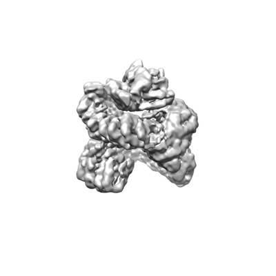 emd_25363.png emd_25363.png | 39.6 KB | ||
| Filedesc metadata | emd-25363.cif.gz | 6.7 KB | ||
| Archive directory |  http://ftp.pdbj.org/pub/emdb/structures/EMD-25363ftp://ftp.pdbj.org/pub/emdb/structures/EMD-25363 http://ftp.pdbj.org/pub/emdb/structures/EMD-25363ftp://ftp.pdbj.org/pub/emdb/structures/EMD-25363 | HTTPS FTP |
-Related structure data
| Related structure data |  7soyMC M: atomic model generated by this map C: citing same article ( |
|---|---|
| Similar structure data |
-Links
| EMDB pages | EMDB (EBI/PDBe) / EMDataResource |
|---|---|
| Related items in Molecule of the Month |


-Map
| File | Download / File: emd_25363.map.gz / Format: CCP4 / Size: 421.9 MB / Type: IMAGE STORED AS FLOATING POINT NUMBER (4 BYTES) | ||||||||||||||||||||||||||||||||||||
|---|---|---|---|---|---|---|---|---|---|---|---|---|---|---|---|---|---|---|---|---|---|---|---|---|---|---|---|---|---|---|---|---|---|---|---|---|---|
| Projections & slices | Image control
Images are generated by Spider. | ||||||||||||||||||||||||||||||||||||
| Voxel size | X=Y=Z: 0.53 Å | ||||||||||||||||||||||||||||||||||||
| Density |
| ||||||||||||||||||||||||||||||||||||
| Symmetry | Space group: 1 | ||||||||||||||||||||||||||||||||||||
| Details | EMDB XML:
|
 Z (Sec.)
 Y (Row.)
 X (Col.)


-Supplemental data
- Sample components
-Entire : The structure of PP2A-B56gamma holoenzyme-PME-1 complex
| Entire | Name: The structure of PP2A-B56gamma holoenzyme-PME-1 complex |
|---|---|
| Components |
|
-Supramolecule #1: The structure of PP2A-B56gamma holoenzyme-PME-1 complex
| Supramolecule | Name: The structure of PP2A-B56gamma holoenzyme-PME-1 complex type: complex / ID: 1 / Parent: 0 / Macromolecule list: all |
|---|---|
| Source (natural) | Organism: Homo sapiens (human) |
-Macromolecule #1: Serine/threonine-protein phosphatase 2A 65 kDa regulatory subunit...
| Macromolecule | Name: Serine/threonine-protein phosphatase 2A 65 kDa regulatory subunit A alpha isoform type: protein_or_peptide / ID: 1 / Number of copies: 1 / Enantiomer: LEVO |
|---|---|
| Source (natural) | Organism: Homo sapiens (human) |
| Molecular weight | Theoretical: 65.378344 KDa |
| Recombinant expression | Organism:  |
| Sequence | String: MAAADGDDSL YPIAVLIDEL RNEDVQLRLN SIKKLSTIAL ALGVERTRSE LLPFLTDTIY DEDEVLLALA EQLGTFTTLV GGPEYVHCL LPPLESLATV EETVVRDKAV ESLRAISHEH SPSDLEAHFV PLVKRLAGGD WFTSRTSACG LFSVCYPRVS S AVKAELRQ ...String: MAAADGDDSL YPIAVLIDEL RNEDVQLRLN SIKKLSTIAL ALGVERTRSE LLPFLTDTIY DEDEVLLALA EQLGTFTTLV GGPEYVHCL LPPLESLATV EETVVRDKAV ESLRAISHEH SPSDLEAHFV PLVKRLAGGD WFTSRTSACG LFSVCYPRVS S AVKAELRQ YFRNLCSDDT PMVRRAAASK LGEFAKVLEL DNVKSEIIPM FSNLASDEQD SVRLLAVEAC VNIAQLLPQE DL EALVMPT LRQAAEDKSW RVRYMVADKF TELQKAVGPE ITKTDLVPAF QNLMKDCEAE VRAAASHKVK EFCENLSADC REN VIMSQI LPCIKELVSD ANQHVKSALA SVIMGLSPIL GKDNTIEHLL PLFLAQLKDE CPEVRLNIIS NLDCVNEVIG IRQL SQSLL PAIVELAEDA KWRVRLAIIE YMPLLAGQLG VEFFDEKLNS LCMAWLVDHV YAIREAATSN LKKLVEKFGK EWAHA TIIP KVLAMSGDPN YLHRMTTLFC INVLSEVCGQ DITTKHMLPT VLRMAGDPVA NVRFNVAKSL QKIGPILDNS TLQSEV KPI LEKLTQDQDV DVKYFAQEAL TVLSLA UniProtKB: Serine/threonine-protein phosphatase 2A 65 kDa regulatory subunit A alpha isoform |
-Macromolecule #2: Isoform Gamma-1 of Serine/threonine-protein phosphatase 2A 56 kDa...
| Macromolecule | Name: Isoform Gamma-1 of Serine/threonine-protein phosphatase 2A 56 kDa regulatory subunit gamma isoform type: protein_or_peptide / ID: 2 / Number of copies: 1 / Enantiomer: LEVO |
|---|---|
| Source (natural) | Organism: Homo sapiens (human) |
| Molecular weight | Theoretical: 52.69527 KDa |
| Recombinant expression | Organism: |
| Sequence | String: MLTCNKAGSR MVVDAANSNG PFQPVVLLHI RDVPPADQEK LFIQKLRQCC VLFDFVSDPL SDLKWKEVKR AALSEMVEYI THNRNVITE PIYPEVVHMF AVNMFRTLPP SSNPTGAEFD PEEDEPTLEA AWPHLQLVYE FFLRFLESPD FQPNIAKKYI D QKFVLQLL ...String: MLTCNKAGSR MVVDAANSNG PFQPVVLLHI RDVPPADQEK LFIQKLRQCC VLFDFVSDPL SDLKWKEVKR AALSEMVEYI THNRNVITE PIYPEVVHMF AVNMFRTLPP SSNPTGAEFD PEEDEPTLEA AWPHLQLVYE FFLRFLESPD FQPNIAKKYI D QKFVLQLL ELFDSEDPRE RDFLKTTLHR IYGKFLGLRA YIRKQINNIF YRFIYETEHH NGIAELLEIL GSIINGFALP LK EEHKIFL LKVLLPLHKV KSLSVYHPQL AYCVVQFLEK DSTLTEPVVM ALLKYWPKTH SPKEVMFLNE LEEILDVIEP SEF VKIMEP LFRQLAKCVS SPHFQVAERA LYYWNNEYIM SLISDNAAKI LPIMFPSLYR NSKTHWNKTI HGLIYNALKL FMEM NQKLF DDCTQQFKAE KLKEKLKMKE REEAWVKIEN LAKANPQVLK KRIT UniProtKB: Serine/threonine-protein phosphatase 2A 56 kDa regulatory subunit gamma isoform |
-Macromolecule #3: Serine/threonine-protein phosphatase 2A catalytic subunit alpha i...
| Macromolecule | Name: Serine/threonine-protein phosphatase 2A catalytic subunit alpha isoform type: protein_or_peptide / ID: 3 / Number of copies: 1 / Enantiomer: LEVO / EC number: protein-serine/threonine phosphatase |
|---|---|
| Source (natural) | Organism: Homo sapiens (human) |
| Molecular weight | Theoretical: 35.636152 KDa |
| Recombinant expression | Organism:  Trichoplusia ni (cabbage looper) Trichoplusia ni (cabbage looper) |
| Sequence | String: MDEKVFTKEL DQWIEQLNEC KQLSESQVKS LCEKAKEILT KESNVQEVRC PVTVCGDVHG QFHDLMELFR IGGKSPDTNY LFMGDYVDR GYYSVETVTL LVALKVRYRE RITILRGNHE SRQITQVYGF YDECLRKYGN ANVWKYFTDL FDYLPLTALV D GQIFCLHG ...String: MDEKVFTKEL DQWIEQLNEC KQLSESQVKS LCEKAKEILT KESNVQEVRC PVTVCGDVHG QFHDLMELFR IGGKSPDTNY LFMGDYVDR GYYSVETVTL LVALKVRYRE RITILRGNHE SRQITQVYGF YDECLRKYGN ANVWKYFTDL FDYLPLTALV D GQIFCLHG GLSPSIDTLD HIRALDRLQE VPHEGPMCDL LWSDPDDRGG WGISPRGAGY TFGQDISETF NHANGLTLVS RA HQLVMEG YNWCHDRNVV TIFSAPNYCY RCGNQAAIME LDDTLKYSFL QFDPAPRRGE PHVTRRTPDY FL UniProtKB: Serine/threonine-protein phosphatase 2A catalytic subunit alpha isoform |
-Macromolecule #4: Protein phosphatase methylesterase 1
| Macromolecule | Name: Protein phosphatase methylesterase 1 / type: protein_or_peptide / ID: 4 / Number of copies: 1 / Enantiomer: LEVO / EC number: protein phosphatase methylesterase-1 |
|---|---|
| Source (natural) | Organism: Homo sapiens (human) |
| Molecular weight | Theoretical: 42.352379 KDa |
| Recombinant expression | Organism: |
| Sequence | String: MSALEKSMHL GRLPSRPPLP GSGGSQSGAK MRMGPGRKRD FSPVPWSQYF ESMEDVEVEN ETGKDTFRVY KSGSEGPVLL LLHGGGHSA LSWAVFTAAI ISRVQCRIVA LDLRSHGETK VKNPEDLSAE TMAKDVGNVV EAMYGDLPPP IMLIGHAMGG A IAVHTASS ...String: MSALEKSMHL GRLPSRPPLP GSGGSQSGAK MRMGPGRKRD FSPVPWSQYF ESMEDVEVEN ETGKDTFRVY KSGSEGPVLL LLHGGGHSA LSWAVFTAAI ISRVQCRIVA LDLRSHGETK VKNPEDLSAE TMAKDVGNVV EAMYGDLPPP IMLIGHAMGG A IAVHTASS NLVPSLLGLC MIDVVEGTAM DALNSMQNFL RGRPKTFKSL ENAIEWSVKS GQIRNLESAR VSMVGQVKQC EG ITSPEGS KSIVEGIIEE EEEDEEGSES ISKRKKEDDM ETKKDHPYTW RIELAKTEKY WDGWFRGLSN LFLSCPIPKL LLL AGVDRL DKDLTIGQMQ GKFQMQVLPQ CGHAVHEDAP DKVAEAVATF LIRHRFAEPI GGFQCVFPGC UniProtKB: Protein phosphatase methylesterase 1 |
-Experimental details
-Structure determination
| Method | cryo EM |
|---|---|
 Processing Processing | single particle reconstruction |
| Aggregation state | particle |
-Sample preparation
| Concentration | 1 mg/mL |
|---|---|
| Buffer | pH: 8 |
| Grid | Model: Quantifoil R1.2/1.3 / Material: COPPER / Mesh: 300 / Pretreatment - Type: GLOW DISCHARGE |
| Vitrification | Cryogen name: ETHANE |
- Electron microscopy
| Microscope | FEI TITAN KRIOS |
|---|---|
| Image recording | Film or detector model: GATAN K3 BIOQUANTUM (6k x 4k) / Average electron dose: 50.8 e/Å2 |
| Electron beam | Acceleration voltage: 300 kV / Electron source:  FIELD EMISSION GUN FIELD EMISSION GUN |
| Electron optics | Illumination mode: OTHER / Imaging mode: BRIGHT FIELD / Nominal defocus max: 2.3000000000000003 µm / Nominal defocus min: 1.5 µm |
| Experimental equipment |  Model: Titan Krios / Image courtesy: FEI Company |
-Image processing
| Startup model | Type of model: OTHER / Details: Ab initio model generated by cryoSparc |
|---|---|
| Final reconstruction | Resolution.type: BY AUTHOR / Resolution: 3.4 Å / Resolution method: FSC 0.143 CUT-OFF / Number images used: 276737 |
| Initial angle assignment | Type: MAXIMUM LIKELIHOOD |
| Final angle assignment | Type: MAXIMUM LIKELIHOOD |
